Contact dermatitis caused by ambulatory blood pressure monitoring
Article information
A 63-year-old housewife who had diabetes came to the outpatient clinic for evaluation of labile hypertension. She had undergone a total thyroidectomy 8 years earlier to treat papillary thyroid cancer. She had no history of allergy to any medicine or food. Twenty-four-hour ambulatory blood pressure monitoring was done. After this test, she developed a pruritic erythematous patch on her upper arm, which had been in contact with the blood pressure cuff (Fig. 1). Contact dermatitis after ambulatory blood pressure monitoring has not been reported. She complained markedly about this unexplained complication, which improved after treatment with antihistamines and topical steroid cream.
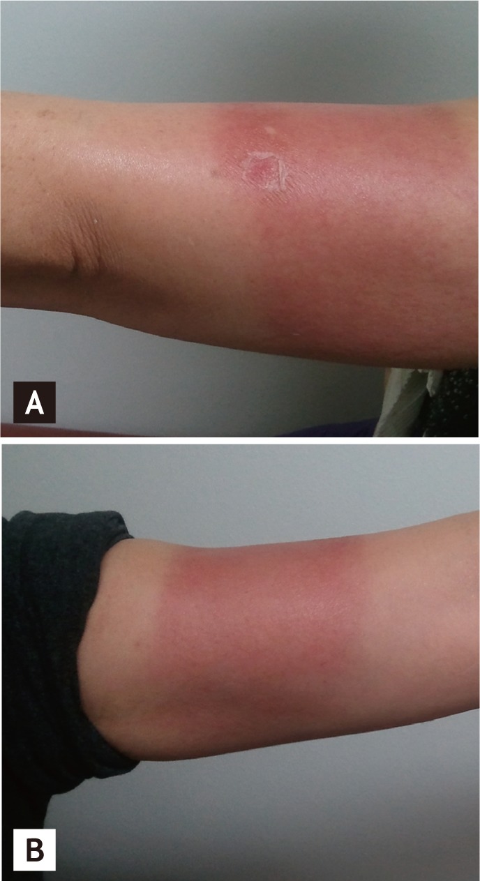
Pruritic erythematous patch on upper arm. (A) Lateral side. (B) Medial side.
Contact dermatitis, a common problem in clinical practice, is defined as an inflammatory reaction in response to substances that come in contact with the skin. There are multiple risk factors for developing contact dermatitis. Extrinsic factors include low-humidity, hot, or occlusive environments and occupations that decrease the protective barrier of the skin, including hairdressers, construction workers, health care workers, agricultural workers, and cleaners. Intrinsic factors include Asian skin, which is more reactive than white or black skin, female sex, older age, and a history of atopic dermatitis. We suggest a careful explanation of this possible effect from a common clinical test, especially to patients with risk factors for contact dermatitis.
Notes
No potential conflict of interest relevant to this article is reported.