Biopsy-Proven Type 1 Renal Tubular Acidosis in a Patient with Metabolic Acidosis
Article information
A 44-year-old female was referred to our outpatient clinic with metabolic acidosis and vomiting. She had a history of toxic hepatitis, but no medications. On admission, her blood pressure was 120/70 mmHg with a pulse of 68 beats/min. The laboratory findings showed normal anion gap metabolic acidosis: Na 140 mmol/L, K 1.4 mmol/L, Cl 113 mmol/L, pH 7.192, and bicarbonate 12 mmol/L. The urine chemistry was pH 7.0, Na 55 mmol/L, K 14 mmol/L, and Cl 63 mmol/L (hyperchloremic metabolic acidosis of renal origin). Abdominal computed tomography revealed medullary nephrocalcinosis (Fig. 1). The renal pathology was non-specific. There was decreased net H+ secretion in the collecting tubules, resulting in a high urine pH. Acidification in the collecting tubules is primarily achieved via H secretion by luminal H+-ATPase. However, immunostaining for H+-ATPase was negative (Fig. 2). After alkali replacement with sodium bicarbonate, the serum bicarbonate recovered to above the lower normal limit (26.6 mmol/L). She was discharged in an improved condition.
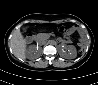
Non-contrast computed tomography shows medullary nephrocalcinosis (arrows).

Immunostaining for aquaporin-2 and H+-ATPase in patient kidney (× 100). (A) Immunostaining for aquaporin-2 in principle cells of collecting tubule (arrows). (B) No immunostaining were observed in the collecting tubule (open arrows).
Notes
No potential conflict of interest relevant to this article was reported.