A Tulip-Shaped Gastric Carcinoid Tumor
Article information
A 44-year-old woman was referred for endoscopic ultrasound (EUS) evaluation of a gastric submucosal tumor that was found during a health checkup. At endoscopy, a tulip-like mass with a central lobulated dent was found in the upper body of the stomach (Fig. 1). A biopsy disclosed only dilated blood vessels in edematous stroma. EUS revealed a 29 × 25-mm inhomogeneously hypoechoic protruding mass in the submucosal layer (Fig. 2). Contrast-enhanced computed tomography revealed a high-enhancing submucosal mass in the stomach with no metastatic lesions. A laparoscopic wedge resection was done. The resected tumor had ribbon-like patterns of uniform cells with round nuclei and pink cytoplasm, confined mainly to the submucosa (Fig. 3A). Immunohistochemically, the tumor cells were stained with synaptophysin (Fig. 3B). The mass was diagnosed as a gastric carcinoid tumor. This patient has been followed for 2 years with no evidence of recurrence.
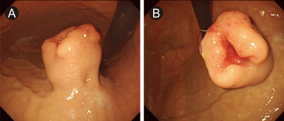
At endoscopy, a smooth, tulip-like mass with a central lobulated dent was found in the upper body of the stomach (A, B).
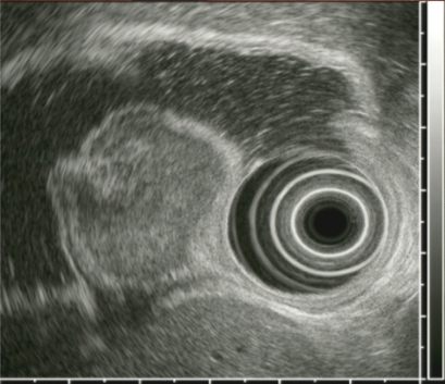
Endoscopic ultrasound revealed an about 29 × 25-mm inhomogeneously hypoechoic protruding mass in the submucosal layer.
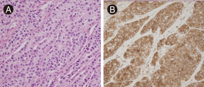
(A) Light microscopy showed ribbon-like patterns of uniform cells with round nuclei and pink cytoplasm, mainly confined to the submucosa (H&E, × 100). (B) On immunohistochemical study, the tumor cells were stained with synaptophysin (× 100).
Notes
No potential conflict of interest relevant to this article was reported.