A Simple Quantitative Assay of Vascular Plasminogen Activator:
— Not Only Stimulated Firinolytic Activity But Also Basal Firinolytic Activity are Correlated with Venous Fibrinolytic Activity —
Article information
Abstract
A simple method for the quantitative assay of vascular plasminogen activator is described. From 24 chronic renal failure patients who were undergoing surgery for A/V fistular or shunt construction, samll pieces of artery (mean 7.8 mg, range 3.0 – 18.5 mg) and vein (mean 4.5 mg, range 3.0 – 8.8 mg) were used. The fibrinolytic acitivity was assayed on a well-controlled fibrin plate by only distilled water immersion and incubation at 37°C. Basal fibrinolytic activity was correlated with venous fibrinolytic activity (r = 0.773, p<0.0001) and with arterial fibrinolytic activity (r = 0.55, 0.005<p<0.01). The increment of fibrinolytic activity by venous occlusion was correlated to venous fibrinolytic activity (r = 0.849, p < 0. 0001) but not to arterial fibrinolytic activity (r = 0.34, 0.1<p<0.25).
INTRODUCTION
The methods currently used to measure plasminogen activator (PA) in tissue are the potassium thiocyanate extraction technique1) and the histochemical fibrin slide method, first described by Todd2) and modified by Pandolfi3). Many investigators have studied the relation between disease and tissue PA4–8) using these methods, But despite various scoring systems, they remain semiquantitative ones. Also, it is not easy to obtain a human blood vessel in a physiologic state, so most studies dealing with vascular PA activity have been based on postmortem specimens or major resectional surgery specimens. There is no controversy over the fact that the main factor of fibrinolytic activity, t-PA, is from vascular endothelial cells, especially of the vein. But it is not known whether any correlation exists between the plasminogen activator in the arteries and/or veins and systemic fibrinolytic activity, because the PA activity of the vessels varies from one anatomic region to another4–9,13). To assay the physiological and clinical significance of vascular PA activity, it is necessary to obtain artery and vein specimens in a homogenized condition from the given anatomic location. A/V fistular or shunt operation in chronic renal failure for hemodialysis gives us a good opportunity to obtainan artery or vein from the same anatomical area conveniently. In many cases, especially side-to-side anastomosis, small pieces of artery or vein can be obtained. Unfortunately, the quantity of the material is too small to extract fibrinolytic activity by the previously described methods, so we attempted a new method of extracting fibrinolytic activity from a small piece of vessel. With this new method, we could measure the fibrinolytic activity of a piece of vessel as small as 3 mg of wet weight. We compared vascular fibrinolytic activity with systemic fibrinolytic activity of basal and response to venous occlusion.
MATERIAL AND METHOD
1. Samples of Artery and Vein
Samples of artery and vein were obtained from 24 chronic renal failure patients during internal fistular or external shunt operation between the radial artery and superficial cephalic vein at the forearm for hemodialysis. The distribution of age, sex, and underlying disease of renal failure are shown in Table 1. The material was rinsed in cold saline, debried attached tissue, and weighed in a weighing bottle to prevent weight change due to evaporation during the weighing procedure. Our preliminary study showed that the weight had to be more than 3 mg to be on a dose-dependent curve of fibrinolytic activity and tissue weight. The average weight of the artery was 7.8 mg (range 3.0 – 18. 5 mg) and of the vein 4.46 mg (range 3.0 – 8.8 mg). That was the reason why we could not use dry weight.

Age, Sex and Underlying Disease Distribution of 24 Chronic Renal Failure Patients
2. Plasma Sampling and Venous Occlusion
Blood sampling was done bwtween 8 a.m. and 9 a.m. after overnight fasting. Venous occlusion was produced by application of a sphygmomanometer cuff wrapped round the upper arm and inflated for 10 minutes at 100 mmHg. Blood samples were obtained from an antecubital vein of the same side with fistular and/or shunt operation before application of the cuff and again after venous occlusion. The blood samples obtained before the venous occlusion were taken either with or with out the help of a tourniquet that had been applied for less than 1 minute. The blood was transferred immediately into plastic tubes containing 2.8% sodium citrate in 9.1 blood/anticoagulant ratio. Platelet poor plasma was separated by centrifugation at 4°C for 30 minutes at 2000 g and stored at −70°C.
3. Fibrin Plate and Euglobulin
1) Reagent
Plasminogen-rich bovine fibrinogen, bovine thrombin, and flufenamate furchased from SIGMA.
2) Solution
Plate buffer: contained 0.05 mol/L sodium diethyl barbiturate, 0.093 mol/L NaCl, 1.66 mmol/ L MgCl2 and was adjusted to PH 7.8 with HCl, ionic strength 0.15
EDTA buffer: contained 0.05 mol/L sodium diethyl barbiturate, 0.10 mol/NaCl, 0.25% (wt/vol) gelatin, and 2.7 mmol/L EDTA and was adjusted to PH 7.8 with HCl, ionic strength 0.15
Dextran sulfate solution: dextran sulfate, 100 mg/l, was dissolved in distilled water.
Sodium flufenamate solution: Flufenamic acid, 0.5 mol/L, was dissolved in an equivalent concentration of sodium hydroxide and diluted to 14 mmol/L with EDTA buffer.
4. Preparation of Euglobulin
0.5 ml plasma was diluted with 4.0 ml cold distilled water, and 0.5 mldextran sulfate solution was added. It was mixed and placed on ice and the PH adjusted to 5.9 (5.85 – 5.9) with acetic acid solution with constant stirring motion. It was left to stand on ice for 30 – 60 minutes, then centrifuged at 2000 g for 10 minutes at 4°C. The supernatant was decanted completely, and the interior of the tube was dried with absorbent tissue. The precipitate was dissolved in 0.5 ml saline barbital buffer.
5. Assay of Tissue Fibrinolytic Activity
The artery and/or vein was placed on a single fibrin plate and overlaid with 30 μl of distilled water, most of which covered the tissue within the global water drop.
6. Assay of Plasma total fibrinolytic Activity
We described before the fibrin plate method of euglobulin fibrinolytic activity14). In brief, three 30 μl drops of dextran sulfate euglobulin fraction were placed onto the surface of the fibrin layer in a triangular fashion. To each drop, 5 μl of sodium flufenamate solution was added directly. Two perpendicular diameters of each lysed zone were measured in mm2 after 17 hours incubation at 37°C.
7. Interpretation
1) Plasma Fibrinolytic Activity
Using the dose-dependent curve of normal pooled plasma, the final result in the blood activator units was compated.
2) Tissue Fibrinolytic Activity
The lysed area (mm2) divided by mg of tissue and expressed as mm2/mg of artery and vein.
RESULTS
1. Dose Curve of Vascular fibrinolytic Activity
A relatively long length of radial artery from one patient was used for the dose-dependent curve. The tissue was resected into various sizes. The weights were 22.4 mg, 12.5 mg, 8.5 mg, and 1.9 mg, retrospectively, and the fibrinolytic activity from each sample was 772 mm2, 475 mm2, 239 mm2, 157 mm2, and 158 mm2, in that order. So the tissues between 3.0 mg and 22.4 mg correlated well with their fibrinolytic activity as measured on the fibrin plate (r = 1, p <0.0001), and the mean of fibrinolytic activity, divided by weight was 39.7 ± 4.4. The fibrinolytic activity of samples less than 3 mg of wet weight was not on the linear dose curve (Fig. 1).
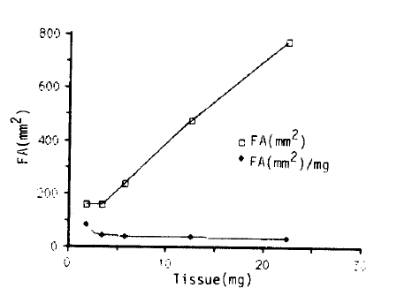
Dose-dependent curve of vein fibrinolytic activity. Fibrinolytic areas were plotted according to various weights of wet vein from the same material.
2. Systemic Basal Fibrinolytic Activity vs Vascular Fibrinolytic Activity
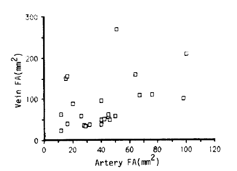
The basal fibrinolytic activity in euglobulin fraction was correlated with the arteral fibrinolytic activity (r = 0.55, 0.005 <p <0.01) anα venous fibrinolytic activity (r = 0.773, p<0.0001) (Fig. 2 A, B).

Correlation between basal morning fibrinolytic activity and artery (A) and vein (B).
3. Increment of Euglobulin Fibrinolytic Activity after Venous Occulsion vs Vascular Fibrinolytic Activity
The increment of fibrinolytic activity after venous occlusion was related to venous fibrinolytic activity (r = 0.849, p <0.0001) but not to arterial fibrinolytic activity (r= 0.338, 0.1 <p <0.25) (Fig. 3 A, B).

Correlation between the incesement of fibrinolytic activity after venous occlusion and artery (A) and vein (B).
DISCUSSION
This study was designed to evaluate the relationship between the fibrinolytic activity and the systemic fibrinolytic activity. Kluft15,16) reports that total fibrinolytic activity in dextran sulfate euglobulin fraction prepared from normal morning plasma mainly consist of factor XII dependent and independent intrinsic fibrinolytic activity, while an additional amount of approximately 1 – 2% is contributed by the extrinsic or vascular activator. However, the physiologic importance and the nature of the intrinsic fibrinolytic activity is uncertain. Our results showing the correlation between the fibrinolytic activity in dextran sulfate euglobulin in the morning, and the fibrinolytic activity of the artery and vein suggest that the intrinsic fibrinolytic activity is a reflection of the fibrinolytic activity of the artery and vein. Vascular fibrinolytic activity is released from endothelial cells by several stimuli17,20), and the amount released is determined by the fibrinolytic capacity or reserve of the endothelial cells21–24).
The fibrinolytic activity of the artery and vein is different24) and varies from one anatomic region to another4,9–13). So it is not easy to characterize the fibrinolytic activity of one branch of vein and/or artery in total fibrinolytic activity of plasma. It was known that tissue plasminogen activator is insoluble under usual conditions. The inability to extract the fibrinolytic agent from tissues with saline was observed by Fleisher25), and Permin26,27) tried unsuccessfully to extract the activator with the following solution: Phosphate buffer (N/15 pH 5.6 – 8.3), acetic acid (1%, 5%), HCl (0.01 N), ammounium hydroxide (0.01 N), sodium hydroxide (0.01 N), and glycerol (87%) containing 0.5% acetic acid. Based on the observation that the tissue fibrinolytic activator extracted from tissue with a strong solution of potassium thiocyanate and with an improved standandized fibrin plate method, a method for the quantitative extraction and assay of the tissue activator was developed by Astrup and Albrechtzen1). Using the plasminogen inactivated fibrin plates, it was demonstrated that the activity present in the extract of tissue was caused by an activator of plasminogen and not by a protease28). But the wide range of activity in the different organs and the large individual variations discouraged consideration of the origin of activator and its correlation with systemic fibrinolytic activity. In our method, fibinolytic activity could be extracted by incubating a small piece of vessel in distilled water dropped on a plasminogen-rich fibrin plate. This method was very simple, sensitive enough to measure a quantity of material as small as 1 mg, and did not require any equipment or reagent except a controlled fibrin plate and distilled water. But the material should be more than 3 mg for two reasons. First, material less than 3 mg is not on a dose-dependent curve, and second, the weight is not reproducible if the weighing material (wet vessel) is less than 3 mg. We do not understand the exact mechanism of how distilled water extracts t-PA. In our preliminary study, the lysed zone was negligible when we used normal saline, half saline, DMEM, or HANK solution instead of distilled water. We examined the vessels microscopically after incubation and found that the endothelium was disrupted and lysed out with distilled water but not with normal saline, half saline, DMEM or HANK solution. So the osmotic lysis of endothelial cells and the release of t-PA might be assumed. Also we speculated that distilled water may change the environment of the released t-PA and its inhibitor. For example, the change of ionic strength might be a factor in separating t-PA from its inhibitor. In conclusion, we could extract vascular fibrinolytic activity by distilled water on a fibrin plate. Basal fibrinolytic activity was linked to artery and venous fibrinolytic activity. The increment of fibrinolytic activity after venous occlusion was linked to venous fibrinolytic activity but not to arterial fibrinolytic activity.