A Case Report: Carcinoma of the Gastric Stump after Billroth II Resection
Article information
Abstract
Carcinoma of the gastric stump as a late complication for resection of benign disease is a well recognized clinical entity.
The risk of stump carcinoma, at least in Western Europe, is estimated to be increased twofold compared with the normal population.
But in Korea there has been one report. Recently we experienced a case of carcinoma of the stump which developed about 20 years after a gastro-jejunostomy for a duodenal ulcer.
INTRODUCTION
Cancer of the gastric stump in patients previously operated on for benign disease was first mentioned by Balfour in 1922 and was subsequently described by several authors. Today, cancer of the gastric stump is no longer the great rarity it was once considered to be. Although the question whether gastric resection for ulcer disease implies an increased risk for subsequent cancer remains a matter of dispute.
Many authors in western Europe agree that there is evidence of increased risk compared with the normal population.
Although stomach cancer is the most common malignancy of adults in Korea, there has been one report of 3 case of stump cancer. So we report a with a review of the literature.
CASE
A 69-year-old man was transferred to our hospital with the chief complaints of hematemesis and melena. About 5 days before admission he had received 5 pints of blood at a private clinic because of hypovolemic symptoms.
He had a history of subtotal gastrectomy 20 years ago due to a bleeding duodenal ulcer. But since then no symptoms related to peptic ulcers had developed till the present.
Two years before admission he checked UGIS at OPD but there was no evidence of a filling defect or ulceration on the remnant stomach and anastomosis.
On physical examination, the patient was pale and presented a chronically ill appearance. His blood pressure was 110/70, pulse rate 110/min, respiration rate 25/min and temperature was 36.5°C, the sclera was not icteric but the conjunctiva was slightly anemic. On abdomen no organomegaly or palpable mass was detected and the surgical scar was noted. All routine laboratory tests were normal with the exception of hemoglobin and hematocrit (8.1 g/dl, 27.1 % respectively), the liver function tests were normal.
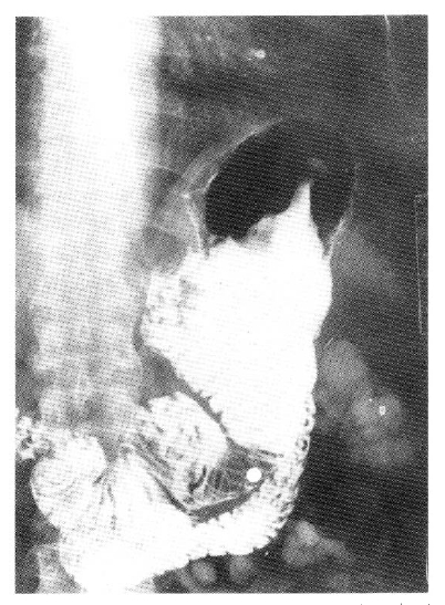
An upper gastrointestinal series revealed a large filling defect with central ulceration, about 6×3 cm in size on the anterior wall side of the residual stomach body (Fig. 1).

A large filling defect with central ulceration about 6×3 cm in size in the anterior wall side of the residual stomach body.
Endoscopy was performed with olympus GIF PIO. A large nodular mass was noted on the anterior wall and lesser curvature aspect of the body 2 cm above the stoma. The mass had superficial ulcerations and was easily bled to the touch. Endoscopic examination of the jejunal side was normal (Fig. 2).

A large nodular mass was noted on the anterior wall an lesser curvature aspect of the body 2 cm above the stoma. The mass had superficial ulcerations and bled easily to the touch. Endoscopic examination of the jejunal side was normal.
The abdominal CT revealed no evidence of regional metastasis to liver, spleen, pancreas and no demonstrable lymphadenopathy in perigastric, retroperitoneal and mesenteric group.
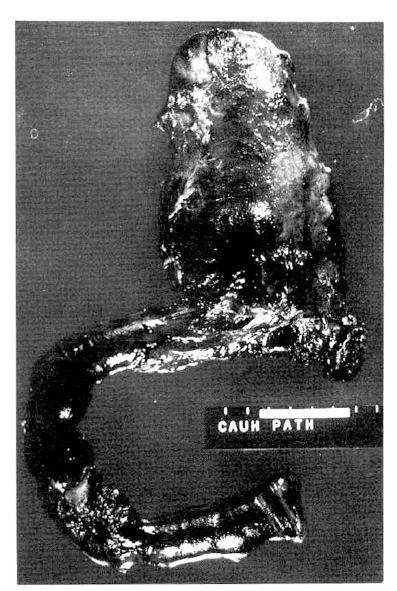
The tumor measured 7.5×5.0 cm in cross diameter and was located 8.5 cm from the proximal resection margin. The tumor did not extend to the jejunum grossly (Fig. 3).

The tumor measure 7.5×5.0 cm in cross diameter and is located 8.5 cm from the proximal resection margin. The tumor does not extend into the jejunum grossly.
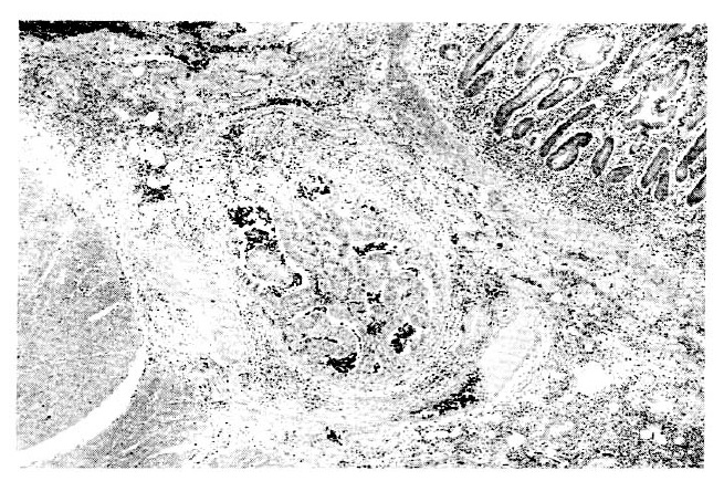
Sections from the gastro-jejunostomy site showed moderately well differentiated adenocarcinoma of the stomach, which directly extended into the anastomosis site of the jejunum (Fig. 4)

Sections from the gastrojejunostomy site show moderately well differentiated adenocarcinoma of the stomach, which is directly extended into the anastomosis site of the jejunum.
Also the tumor was diffusely infiltrated throughout all layers.
DISCUSSION
When defining cancer of the gastric stump, two criteria must be fulfilled; first, the previous surgery (partial gastrectomy or gastroenterostomy) should be for benign disease, and second, to rule out the presence of an occult carcinoma at the time of the surgery a time interval of at least five years should have elapse since the original gastric resection.
Although the interval between resection for ulcer and the appearance of gastric stump cancer varied, it increased to six to eight times as much between the 15th and 30th years after the initial surgery.2) In our case the interval was about 20 years, in 1966 the patient underwent a subtotal gastrectomy due to massive upper GI bleeding.
The incidence of stump cancer varies considerably with geographical location, with a high incidence in certain parts of Scandinavia as well as in central Europe3). Contrary to these findings this form of malignancy is seldom observed in Great Britian or in the United States.4)
In a controlled retrospective study5) of 630 cases of gastric cancer, the frequency of previous gastric surgery for ulcer was increased to about six times more than the control for those patients operated on 25 years previously.
In southern Germany Griesser and Schmit6), reported the frequency of stump cancer was 13.3% among 580 patients after partial gastrectomy for gastric ulcer. Most authors agree to a causal relationship of partial gastrectomy and an increased risk of cancer in the gastric remnant. However some investigators did not observe a higher frequency of stump carcinoma than expected in the normal population with intact stomachs.7,8)
But in Korea, there has been one report of 3 cases of stump cancer, Although stomach cancer is the most common malignancy in adults, stump cancer is very rare. The reasons are not well established, but, one of which is that Billroth II resection was introduced about 30 years ago in this country, therefore, the incidence of stump cancer maybe increase if regular examination has been performel.
Although the etiology of stump cancer has not been determined, clinical and experimental observations have pointed to the important role of the duodenogastic reflux.
Bile, the main component of the doudenogastric reflux is known to act as a detergent thereby damaging the mucosal barrier of the stomach. Atropic gastritis, intestinal metaplasia, and cystification of the mucosal glands are common findings in the vicinity of a gastroenteric anastomsis.
The prominence of a Billroth II anastomosis or a gastroenterostomy rather than a Billroth I gastroduodenal anastomosis undoubtedly reflected the more frequent use of the former procedures in the earlier era.9) However a latter study of 74 patients with a Billroth I anastomosis found stump cancer in 5.4 per cent, suggesting that the risk was present regardless of the type of previous ulcer surgery.
Griesser and Schmidt6), reported the frequency of stump cancer was 13.3% among 580 patients after partial gastrectomy for gastric ulcers, but when the indication for stomach resection was a duodenal ulcer, only 6.25% of the patients developed a cancer. However other investigators reported that there is no difference in the incidence of cancer of the stump as related to the primary ulcer site.
The main problem in stump cancer, as in any gastric cancer, is related to early diagnosis.
Most patients come too late for treatment, because the symptoms are misinterpreted by the patients, and often by their doctors, too, as benign postgastrectomy symptoms.
One should be especially aware of the possibility of cancer in patients, who, after a long symptomless interval, complain of dyspepsia and other nonspecific GI symptoms. In our patient, the X-ray pictures taken 2 years before admission, showed no evidence of malignancy, but had gastroscopy been performed at that time, we could have had different information about the remnant stomach.
X-rays are often difficult to interpret, as benign postoperative changes may be difficult to distinguish from cancer, so gastroscopy is, in our opinion, the best diagnostic method.
Although carcinoma of the gastric stump is either exophytic or appears as an ulcer, exophytic tumors are more common.
The direction of growth is mainly along the minor site of the gastric remnant from distal to proximal. Studies concerning the localization of cancer of the gastric stump have pointed to the stoma as a specific area.10) Hammar11), Studied 65 autopsy specimens and provided strong evidence for a typical site of cancer growth within the Billroth II stoma.
The only treatment for stump cancer as well as gastric cancer, is surgical. From various reports, 20–60% of the patients can be treated by resection12). Saegesser & Waridel13), recommended subtotal gastrectomy by this method, because total gastrectomy is a greater trauma to the patient, and leads to more serious postoperative complications.
The prognosis is poor, but not hopeless. Saegesser & Waridel13) claim that the prognosis in the cases which can be radically operated, is not worse than for operable gastric cancers in general.
CONCLUSION
The etiology of cancer in the gastric stump is still obscure, but the risk of developing cancer increases considerably with increasing time, all patients who have undergone gastric resection should be regularly followed up by gastroscopy.