Mycobacterium Avium Arthritis with Extra-articular Abscess in a Patient with Mixed Connective Tissue Disease
Article information
Abstract
A case of Mycobacterium avium arthritis in a 39-year-old female patient with mixed connective tissue disease (MCTD) was reported. An extra-articular abscess had formed outside the knee joint and extended down the calf. A culture was taken of the abscess and synovial fluid disclosed Mycobacteriun avium. This was resistant to most anti-tuberculosis agents. A combination of anti-tuberculosis drugs followed a total resection of the abscess.
We concluded that M avium septic arthritis could insidiously develop into an extra-articular abscess. A combination of anti-tuberculosis drugs with a total resection of the abscess was an effective treatment.
INTRODUCTION
Infective arthritis due to Mycobacterium avium is rare and most commonly occurs in immunocompromised subjects, such as those receiving immunosuppressive drugs or HIV positive patients1). M avium arthritis is extremely rare in autoimmune diseases. We present a 39-year-old woman with mixed connective tissue disease (MCTD) who developed M avium septic arthritis accompanying an extra-articular abscess in the popliteal fossa and calf.
CASE REPORT
In 1997, the patient was diagnosed with MCTD after developing right knee arthritis, sclerodactly, Raynaud’s phenomenon and digital vasculitis. She took 10 mg of prednisolone after diagnosis of interstitial lung disease in October 1998. In May 2000, the patient received intra-articular injections with triamcinolone in the right knee because of relapsing arthritis. She was admitted to Wallace Memorial Baptist Hospital in September 2001 with right knee pain and swelling, as well as calf swelling.
On admission, her temperature was 37.8°C. Physical examination showed swelling and mild tenderness in the right knee and calf. Both hands also revealed sclerosis with loss of skin flexibility and ulcerations on the tips of her fingers.
On admission, laboratory data was WBC 6,400/mm3, Hb 12.9 g/dL and platelets were 205,500/mm3. At that time, erythrocyte sedimentation rate (ESR) and C-reactive protein (CRP) were 46 mm/h and 1.7 mg/dL. Assays for antinuclear antibody (ANA) with fluorescein-labeled ANA method resulted in high titer (1:5120) and a speckled pattern. Specific autoantibody studies showed a high titer of U1-RNP antibody (3678 IU/mL). None of anti-ds DNA, anti-SS-A/Ro antibody, anti-SS-B/La antibody, anti-Sm antibody, anti-Scl 70 and anti-centromere antibody was positive. Rheumatoid factor was also negative.
Serologic tests for human immunodeficiency virus (HIV) and Venereal Disease Research Laboratory (VDRL) were negative.
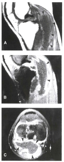
Chest computerized tomography (CT) showed mild esophageal luminal dilatation through the whole length and bilateral subpleural pulmonary fibrosis in both lower lobes of the lungs. Ultrasonography and magnetic resonance image (MRI) around the right knee showed a poorly defined mass lesion laterally to the semi-membranous tendon, anteriorly to the gastrocnemius muscle on the right leg (Figure 1). This extra-articular abscess extended from politeal fossa to the upper end of the Achilles tendon.

Photography shows poorly defined mass lesion at laterally to semi-membranous tendon, anterior to gastrocnemius and along joint space. This mass demonstrates low signal intensity with high signal foci on T1WI/PDWI (A) and patch peripheral enhancement on Gadolium-enhanced T1WI (B, C).
Analysis of the abscess showed decreased viscosity and was yellowish to bloody in color. WBC counts were 700 cells/mm3 and the differential form showed 50% polymorphonuclear leukocyte and 50% lymphocyte. Acid fast bacillus (AFB) stain showed strong positive but polymerase chain reaction (PCR) for mycobacterium tuberculosis was organisms negative. Gram stain was neither positive, nor was growth of on the culture.
Total excision of the abscess was attempted. A small window connected between synovial cavity of the knee and the huge cystic mass included multiple separated rooms. Pathologic findings of the specimen disclosed caseous central necrosis surrounding the granuloma (Figure 2).

Microphotography shows prominent caseous necrosis and surrounding granuloma composed of epitheloid histiocytes and inflammatory cells (big box). The AFB stain shows Mycro-bacterium organisms (arrows, small box).
We initially used the following anti-tuberculosis medications: isoniazide, rifampicin, ethambutol and pyrazinamide. Two months later, the culture for AFB disclosed M. avium in the PCR RFLP assay (PRA) and was resistant against multiple anti-tuberculosis drugs, except cycloserin, in sensitivity testing.
After 9 months of anti-tuberculosis therapy, including cycloserin, we followed up with monthly ultrasonography. Some small cysts persisted between the soleus and the gatrocnemius muscle. We aspirated fluid using a guided ultrasonography. Aspirated fluid was clear of serosangious. Stain for AFB were negative. Culture for Mycobacterium was also negative. We continued the anti-tuberculosis therapy for 12 months and regularly followed up with ultrasonography after discontinuing the medication.
DISCUSSION
This patient with MCTD demonstrated atypical tuberculosis infection in the knee joint. Mycobacterium avium is a rare pathogen in human individuals who have compromised immune systems, such as in AIDS patients. The first reported case of disease due to M avium in a human appeared 50 years ago in a patient with pulmonary involvement2). These infections result from percutaneous inoculation or hematogenous seeding. Cheatum et al. reported the first case of M avium septic arthritis in the knee in 19763). Several case reports and literature reviews were presented4, 5).
Most of the case reports were patients with AIDS. Recently two cases of M avium arthritis in patients with autoimmune disease, such as scleroderma and polymyositis were reported6). This is the first case of M avium septic arthritis and the formation of extra-articular abscess in MCTD. M tuberculosis abscess usually showed to be a thin and smooth-rim enhancement on the MRI7). This case was patch peripheral enhancement on Gadolium-enhanced T1WI.
MCTD is one of overlapping syndrome, such as systemic lupus erythematosus, scleroderma, polymyosite and others, especially associated with a specific antibody namely U1-RNP antibody8). Diagnosis of the MCTD was followed to Alarcon-Segovia and Villareal criteria in this case9).
The reservoirs and routes of the transformation of M avium are unknown. It has been suggested by other authors that microtrauma of the joints or repeated injections of intraarticular steroids may predispose a patient to mycobacterial infection3, 10). In this case, the patient had also had previous intra-articular injections at other clinics.
M avium is highly resistant to multiple anti-tuberculous drugs and often a combination of drugs should be used. In M avium osteomyelitis, it has been recognized that surgical debriment, in addition to chemotherapy, is usually necessary for clinical improvement2, 8). This case showed resistance to most anti-tuberculous drugs, except cycloserin. So we used a combination of anti-tuberculosis agents, including cycloserin, for 12 months after total resection of the abscess and followed up with ultrasonography.
We concluded that MCTD or immunosuppressive drugs may be related to M avium arthritis and it could insidiously develop into an extra-articular abscess. A combination of anti-tuberculosis drugs with total resection of the abscess might be necessary.