


INTRODUCTION
Natural Killer (NK)/T-cell lymphoma is a distinct clinicopathologic entity highly associated with Epstein-Barr virus (EBV)1). The disease has protean clinical features characterized by a destruction of the upper respiratory tract, particularly the nasal cavity, palate and paranasal sinuses3). Pathologically, angiocentric and angiodestructive patterns, marked necrosis and infiltration of atypical lymphoid cells are found in most of the lesions. NK/T-cell lymphoma also has a characteristic immunophenotype: CD2-positive, CD56-positive, but usually surface CD3-negative1). NK/T-cell lymphoma is closely linked to a variety of medical complications, such as hemophagocytic syndrome, sepsis and bleeding. Rarely, patients can develop a second primary cancer during the follow-up period and this can be a cause of death3). We experienced a case of NK/T-cell lymphoma complicated by a squamous cell carcinoma of the hard palate during combination chemotherapy and radiation therapy and report the case with a review of the literature.
CASE
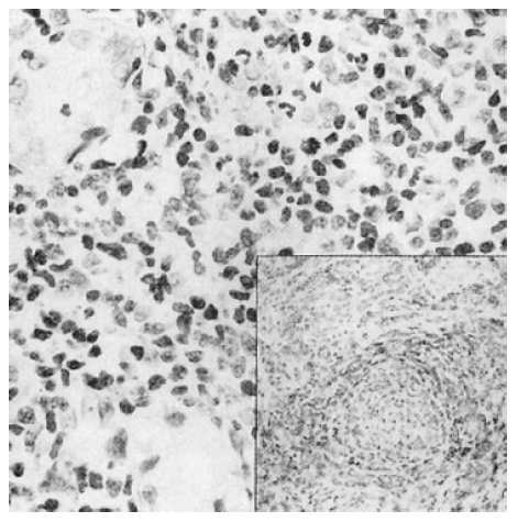
A 50-year-old male patient was admitted to the hospital with complaints of sore throat, fever and bloody sputum for three months. The patient had been suffering from a recurrent sore throat for one year without seeking medical attention. On admission, he had a fever up to 39┬░C mainly at night, but he denied weight loss or night sweating. Physical examination showed multiple lymphadenopathy, including bilateral cervical area along the jugular chain. There was no splenomegaly or hepatomegaly. Diffuse erythematous lesions and wall thickness in the oropharyngeal area were seen on otolaryngeal examination. Laboratory findings were as follows: WBC 7,500/mm3, Hb 12.5 g/dL, Hct 37%, platelet 250,000/mm3, normal urine analysis, ALT/AST 25/40 U/L, serum alkaline phosphatase 95 IU/L and LDH 288 U/L. Chest X-ray finding was normal. A computed tomographic (CT) scan of the neck showed a soft tissue mass in both oropharynx, soft palate and posterior laryngeal wall, and multiple lymph node enlargements of both cervical areas (Figure 1). For a pathologic confirmation, multiple biopsies were performed on the oropharyneal mass and other surrounding areas. Microscopic examination showed an infiltration of atypical small irregularly shaped lymphoid cells admixed with plasma cells, neutrophils and eosinophils. On immunophenotyping study, CD56 and cytoplasmic CD3 were positive and CD20 was negative (Figure 2). According to the microscopic examination and immunophenotype, a diagnosis of NK/T-cell lymphoma was made. For a staging work-up, bilateral bone marrow aspiration and biopsy, gastroscopic examination, bone scan and abdomino-pelvic CT were performed. All of these studies revealed negative findings. The final diagnosis was NK/T-cell lymphoma with stage IIB. The patient was treated initially with combination chemotherapy with COPBLAM-V (cyclophosphamide, vincristine, adriamycin, bleomycin, prednisolone and procarbazine). After 2 cycles of chemotherapy, a follow-up CT scan showed a partial improvement of the mass lesions in the oropharynx. However, after further two cycles of chemotherapy, he developed high fever, severe sore throat and neutropenia. Since an aggravation of the mass lesion was noted on a follow-up CT scan, salvage radiation therapy was given to the patient. Unfortunately, the radiation therapy discontinued due to a sustained neutropenia during the treatment period. The patient was referred to the Hanyang University KURI hospital for further evaluation and treatment.
On admission, the patient looked pale and sick. Physical examination revealed a newly-developed large ulcerative lesion on the hard palate. It seemed that the lesion was developed from the previous radiation therapy. The ulcerative lesion did not heal and rather, increased in size during the hospital stay. Since the new lesion was separated from the initial lymphoma site, we performed a biopsy on the ulcerating lesion of the hard palate. Unexpectedly, the biopsy revealed a moderately differentiated squamous cell carcinoma with a p53 immunoreactivity (Figure 3). The ulcerated lesion was directly connected to the nasal floor. We decided to treat the patient with radiation therapy for the hard palate carcinoma. However, the patient could not tolerate the radiation therapy due to a poor general condition. Although a salvage combination chemotherapy was administered to the patient, a severe hepatic dysfunction and sustained pancytopenia developed without an improvement of the lymphoma or carcinoma lesion and he eventually died of sepsis.
DISCUSSION
The nasal lymphoma expressing a T- or NK-cell phenotype is a disorder with protean clinical features characterized by a destruction of the upper respiratory tract, particularly the nasal cavity, palate and paranasal sinuses3). Pathologically, most of the lesions show angiocentric and angiodestructive patterns, marked necrosis and infiltration of atypical lymphoid cells and are highly associated with EBV. The nasal NK/T-cell lymphoma has a characteristic immunophenotype: CD2-positive, CD56-positive, but usually surface CD3-negative. Cytoplasmic CD3 reactivity can be detected in paraffin sections1,4). Epidemiologically, NK/T-cell lymphoma is rare in the United States and Europe, but relatively common in Asia1,2).
The most important aspect of the nasal NK/T cell lymphoma, as in our case, is that the disease is frequently associated with a variety of medical complications or immunologic disorders3). It has been suggested that the angiocentric lymphomas occasionally retain an immunologic dysfunction, which may be reflected in some clinical manifestations observed in patients with T-lymphocyte malignancies. Ishii et al5) and Sordillo et al6) found a profound immunologic deficit in a few patients with angiocentric lymphomas, probably resulting from the impairment of the T-lymphocyte function. Kim et al3) and Ng et al7) reported that a hemophagocytic syndrome can occur at any time during the clinical course of the disease and is usually rapidly fatal over several weeks. Also, there is a tendency toward an extranodal involvement of sites such as lungs, liver or gastrointestinal tract. More widespread involvement of unusual sites, such as testes or skin, has been occasionally observed.
Some patients develop second primary cancers during the follow-up period. The most striking clinical outcome in such patients is a rapid progression of the second primary cancer after diagnosis3). Despite an aggressive treatment for the second primary cancer, most of the patients die from a rapid deterioration of the cancer within 1 year, which was the case in our patient. Interestingly, our patient developed a squamous cell carcinoma of the hard palate during combination chemotherapy for the NK/T cell lymphoma, with a very short latent period of the development of the second primary cancer. Although it is plausible that the squamous cell carcinoma of the hard palate coexisted initially with the NK/T cell lymphoma, the fact that multiple blind biopsies were performed on the oral cavity, including soft palate, hard palate, oropharynx and pharyngeal wall, at the initial diagnosis argues against this possibility. Furthermore, the hard palate was initially shown to be normal on physical examination, pathological examination, and CT scan. Only limited information is now available regarding the relationship between angiocentric lymphoma and the development of second primary cancers. In an earlier study of 10 patients with midline lethal granuloma by Fauci et al.8) three patients developed second primary cancers and the authors speculated such patients with aberrant immunologic reactions would have an increased propensity for the development of new cancers.
Until recently, there has been no clear consensus on the optimal management for the NK/T cell lymphoma, probably because of the rarity of the tumor and little understanding of its natural history. Despite the inconsistent responses elicited, radiation treatment is still accepted as the treatment of choice for the localized form of angiocentric lymphoma, and achievement of a complete response after radiation treatment is still the most important determinant of favorable survival9,10).
There are several important aspects in our case. Firstly, the patient initially showed multiple systemic failures, which can often be observed in NK/T-cell lymphoma. Although we did not perform a liver biopsy in this patient, unexplained severe hepatic dysfunction with hyperbilirubinemia and prolonged pancytopenia, even after the withdrawal of chemotherapy, suggested that the patient would have a hemophagocytic syndrome and liver involvement by the lymphoma. Secondly, the patient developed a second primary cancer during the combination chemotherapy and radiation therapy. Since the full dose of chemotherapy or radiation therapy was not administered to the patient and the latent period of the development of the second primary malignancy was very short, the exact mechanism should be elucidated. Finally, although radiation therapy is regarded as the standard treatment for the patients with NK/T cell lymphoma, a large proportion of these patients are frequently associated with a systemic involvement by lymphoma and, therefore, need a multimodality treatment, including more effective chemotherapeutic agents. Also, the development of new drugs specifically targeted against the NK/T lymphoma cells is needed.
In summary, we experienced a case of NK/T-cell lymphoma complicated by a squamous cell carcinoma of the hard palate during combination chemotherapy and radiation therapy.


 PDF Links
 PubReader
 ePub Link
 Full text via DOI
 Download Citation
 Print
Print


