


Introduction
Scleritis or episcleritis may be a harbinger of systemic diseases. While episcleritis is relatively benign, the least severe form of disease, necrotizing scleritis is the most severe disease that can rapidly blind the patient1,2). Although episcleritis may be seen in patients with rheumatic disease, most patients with episcleritis have no underlying connective tissue disease. However, approximately half of patients with scleritis have systemic connective tissue diseases or vasculitic diseases1,3).
RA is the most common rheumatic disease associated with scleritis, followed by WegenerŌĆÖs granulomatosis and relapsing polychondritis. Other less frequently associated diseases include systemic lupus erythematosus, polyarteritis nodosa, ReiterŌĆÖs syndrome, psoriatic arthritis, giant cell arteritis and Cogan syndrome1,4ŌĆō7). The scleritis occurred in patients with BD has been rarely reported in the literature1,8). We describe a female patient who developed the scleritis in the course of BD.
CASE
In December 1999, 46-year-old woman presented with bilateral red eye and boring ocular pain with radiation to the temple developed 2 weeks ago. She had been diagnosed as BehcetŌĆÖs disease in March 1997 by recurrent orogenital ulcerations, pustular skin eruption, erythema nodosum-like lesions and positive pathergy test. She had been admitted in April 1998 to Asan-Kangnung Hospital with bloody diarrhea due to multiple ulcerations of terminal ileum, cecum and ascending colon. At that time, the diagnosis of intestinal BD was established, and prednisolone, colchicine and sulfasalazine were prescribed. Even though taking medications of low dose prednisolone, sulfasalazine, nabumetone and colchicine, she had been suffering from refractory oligoarticular arthritis since July 1999. Her arthritis had been improved with the addition of weekly methotrexate. Recently, she withheld the medications for 6 weeks by herself.
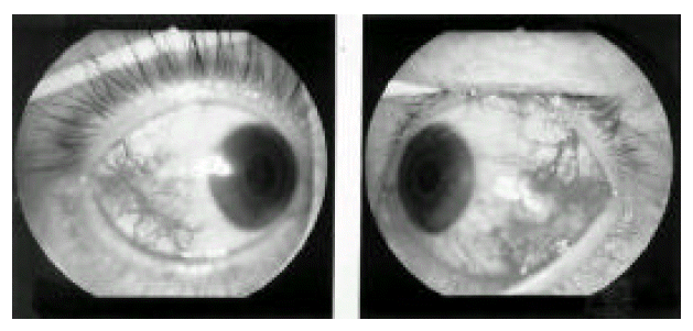
Physical examination revealed multiple aphthous ulcers on the buccal mucosa, several pustular eruptions on the cheek, and multiple erythema nodosum-like lesions on the legs. There were tenderness, swelling and warmth on the right wrist and right knee. On eye examination by an ophthalmologist, there were episcleral nodules with marked dilatation of the scleral vessels and episcleral vessels in both eyes (Figure 1). The vision of both eyes was 20/20. The adjacent cornea and anterior chamber were normal. The focal tenderness with digital compression on both eyes was remarkable.
Hematological and biochemical tests were as follows: WBC 14,300/mm3, hematocrit 25.8 %, platelet 59,8000/mm3, total protein 8.6 g/dL, albumin 3.5 g/dL, AST/ALT 11/6 IU/L. Erythrocyte sedimentation rate was 104 mm/hr and C-reactive protein was 5.51 mg/dL. Antinuclear antibody and rheumatoid factor were negative. Antineutrophil cytoplasmic antibody (ANCA) with p-type was positive (titer, 1:160). HBs antigen and HCV antibody were negative. HLA-B51 antigen was positive. There were no bony abnormalities on plain radiographs of both wrists and both knees.
She was diagnosed to have BD with nodular scleritis. Initially, sulindac 400 mg/day, sulfasalazine 2 g/day, methotrexate 17.5 mg/week and prednisolone 30 mg/day were prescribed. Although her eye disease and arthritis were improved within several days, she experienced two more recurrent events of scleritis when prednisolone was trying to be reduced. After cyclophosphamide was given instead of methotrexate, the scleritis was controlled without ocular complications.
DISCUSSION
Scleritis is a severe, destructive disease that may cause decrease in vision by leading to one or more ocular complications such as anterior uveitis, keratitis, glaucoma, secondary cataract or ocular change1). It is classified clinically as either anterior or posterior scleritis. Anterior scleritis is further classified as either diffuse, nodular or necrotizing scleritis9). Diffuse scleritis is the most common form of scleritis with or without RA. Nodular scleritis is characterized by firm foci of inflammation, which are very tender. Of these three forms of anterior scleritis, necrotizing scleritis is the most destructive form.
Episcleritis is a benign disease that rarely causes decrease in vision. Patients with episcleritis usually describe discomfort rather than pain. The main feature differentiating episcleritis from scleritis is pain, which in the latter is usually severe and deep-seated. Pain is frequently radiated to the temple, brow, sinuses or jaw. Scleritis tends to have recurrences, which can be minimized if the initial attack is adequately treated5). Our patient satisfied the diagnostic criteria by International Study Group for BehcetŌĆÖs Disease10), and she had nodular scleritis with severe boring pain and ocular tenderness. Although she had severe arthritis, the diagnosis of RA could not be established because of asymmetric oligoarthritis, negative rheumatoid factor, no radiographic findings consistent with RA, and her typical symptoms and signs were compatible to BD.
The detection of systemic vasculitic diseases in patients with scleritis can be a sign of poor prognosis. Sainz de la Maza et al11) found that ocular prognosis of scleritis with systemic vasculitic diseases varied depending on the specific systemic vasculitic diseases. Scleritis in spondyloarthropathies or in systemic lupus erythematosus was usually a benign and self-limiting condition, whereas scleritis in WegenerŌĆÖs granulomatosis was a severe disease that could lead to permanent blindness, and that in RA or relapsing polychondritis was a disease of intermediate severity.
The treatment of scleritis requires attention to relief of pain and to halting the progression of the disease. In patients with diffuse and nodular scleritis, NSAIDs should be the initial choice and, in case of therapeutic failure, corticosteroids should be added or substituted as second-line therapy. In patients with necrotizing scleritis, or with diffuse or nodular scleritis which donot respond to NSAIDs and corticosteroids, immunosuppressive agents should be added12). Cyclophosphamide is particularly indicated in ANCA-positive vasculitis-associated scleritis5). Low-dose cyclosporin A has been advised in young patients for whom cyclophosphamide is not recommended because of the risk of sterility13). In our patient with ANCA-positive scleritis, medications of prednisolone, NSAID and low-dose methotrexate were initially given for scleritis and severe arthritis. However, cyclophosphamide was eventually required to control the scleritis.
RA is the most common systemic disease associated with scleritis. The incidence of scleritis in patients with RA is reported in the range of 0.67 to 6.3 percent, although as many as 33 % of all patients presenting to an ophthalmologist with scleritis may have associated RA. The patients with scleritis associated with RA usually have a higher proportion of longstanding and severe erosive joint disease, generalized vasculitis and mortality than those with RA without scleritis14). Scleritis is a manifestation of vasculitis. The scleritis could be the first manifestation of a systemic connective tissue disease or vasculitic disease in considerable patients with scleritis1). As we know, the underlying pathology of BD is vasculitis. Our patient had relatively severe BD with gastrointestinal hemorrhage due to multiple intestinal ulcerations and severe arthritis. It may be speculated that the scleritis might be complicated in severe BD like scleritis in patients with RA. More studies and experiences will be needed.
 PDF Links
 PubReader
 ePub Link
 Full text via DOI
 Download Citation
 Print
Print


