


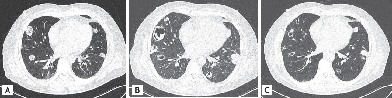
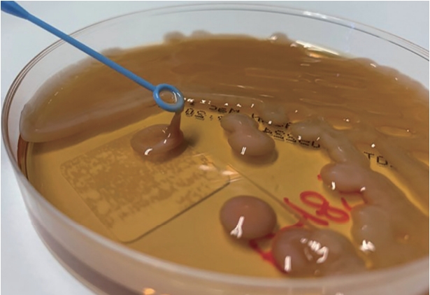
A previously healthy 43-year-old male patient was admitted to our hospital with a 3-day history of pain in the chest and the left scapular area. On admission, the patient was afebrile and had proteinuria. The serum creatinine level was 9.08 mg/dL (normal range, 0.9 to 1.3), and the C-reactive protein level was 27.86 mg/dL (normal range, 0.0 to 0.3). Chest computed tomography (CT) scan revealed multiple mixed ground-glass attenuation nodules of various sizes and masses in both the lungs (Fig. 1A). Abdominal CT scan revealed an 1.7 cm sized enhancing lesion in the S5 region of the liver. Our initial diagnosis was hematogenous pulmonary and liver metastasis or vasculitis-associated glomerulonephritis. However, BacT/Alert blood culture after 5 days revealed the presence of Klebsiella pneumoniae of a hypermucoviscous phenotype, defined by a positive ŌĆ£stringŌĆØ test (Fig. 2). We confirmed a K2 serotype using 16S rRNA gene analysis. The antibiotic regimen was changed to intravenous amikacin and ciprofloxacin. After 1 week, the repeat chest CT scan revealed a marked increase in the extent of multiple necrotic cavitary masses and nodules in both the lungs along with liver abscesses (Fig. 1B). However, ophthalmological examination, brain magnetic resonance imaging, and transthoracic echocardiography, showed no abnormal findings. After 6 weeks, the chest CT scan revealed markedly reduced lesions in both the lungs (Fig. 1C). The antibiotic regimen was de-escalated to ciprofloxacin for 2 months, following which a complete recovery was achieved. Informed consent was obtained from the patient.
A new hypervirulent variant of K. pneumoniae (hvKP) has emerged in Asia. The defining clinical features are the ability to cause serious life-threatening community-acquired infections, including liver abscess, pneumonia, meningitis, and endophthalmitis, and the ability to spread like metastasis in healthy young hosts. Besides, early diagnosis may be delayed in many cases. Herein, we report a case of hvKP infection initially misdiagnosed as malignant metastasis or vasculitis in a healthy adult.
 PDF Links
 PubReader
 ePub Link
 Full text via DOI
 Download Citation
 Print
Print


