


To the Editor,
Interferon-╬▒ (IFN╬▒) is a cytokine with multiple activities including antiviral and anti-proliferative effects, and its various forms including recombinant interferons, purified natural leukocytes, and lymphoblastoid interferons have been widely used to treat hepatitis C virus (HCV) infection [1,2]. The pegylated form, pegylated IFN╬▒ (Peg-IFN╬▒), is used as a once weekly regimen for its convenience to treat HCV infection. Still, its side effects such as flu-like symptoms, mood changes, insomnia, diarrhea, nausea, pruritis, alopecia, and changes in thyroid function or complete blood cell counts are obstacles in maintaining the treatment [1-3].
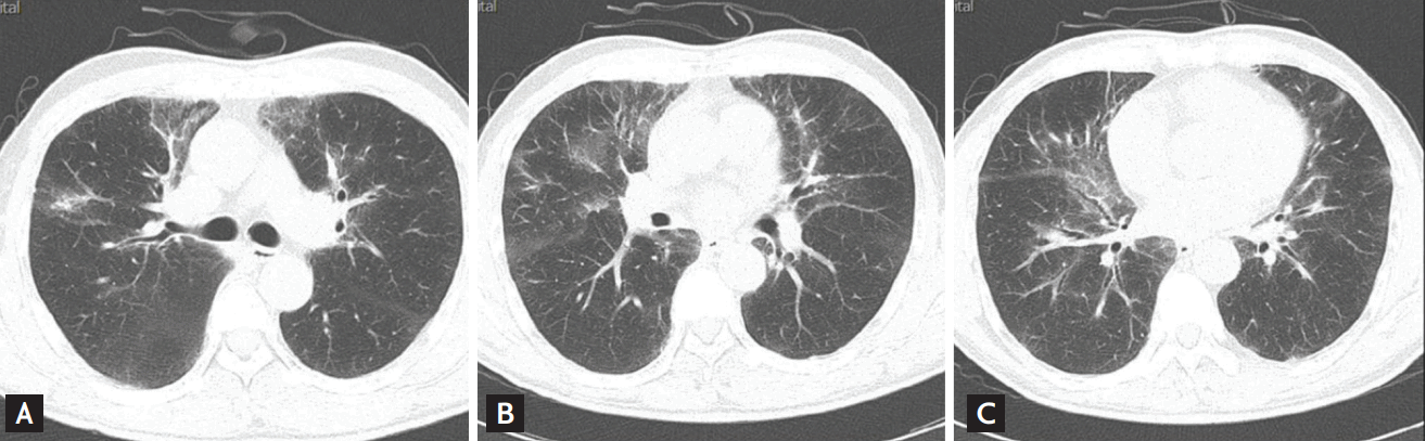
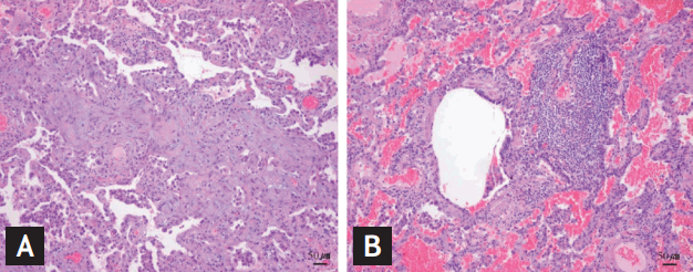
A 52-year-old Chinese male patient visited our hepatology clinic for further evaluation of abnormal liver functions in blood test and fatty changes of the liver parenchyma in ultrasonography, which were incidentally found at another medical institution during a periodic medical checkup 6 months prior to the visit. Laboratory tests revealed anti-HCV antibody to be positive. Serum aspartate aminotransferase and alanine aminotransferase was elevated to 48 and 94 U/L, respectively, and triglyceride elevated to 259 mg/dL. With an impression of HCV infection, further serologic markers were checked. Real-time quantitative HCV-RNA polymerase chain reaction resulted in HCV RNA titer to be 58,500 IU/mL, and its genotype was 2 a/c. Since there were no other definite abnormal findings in physical exam or blood tests including complete blood cell count, thyroid function test or tumor markers, standard treatment regimen using Peg-IFN╬▒2b (80 ╬╝g) injection once weekly along with ribavirin (800 mg) as daily dose was planned for 24 weeks. On the 10th week, the patient complained of some cough. There were no specific changes in physical exam, and the medication was kept on since the symptoms were quite tolerable. Two weeks after, with a total treatment of 13 injections of Peg-IFN╬▒2b, the dry cough still remained, and chest X-ray and computed tomography (CT) for the lung field was done. Some changes were observed in the imaging studies. Chest X-ray showed irregular, linear, and reticular densities involving both central peribronchovascular regions in middle and lower lung fields while there were no definite abnormal findings in the X-ray taken previous to the treatment (Fig. 1A and 1B), and chest CT revealed smooth interlobular septal thickenings with mild ground glass opacity in anterior and medial subpleural regions of both upper lobes, almost entire parenchyma of right middle lobe, left lingular segment, and both lower lobes suggesting subacute phase of interstitial lung disease (Fig. 2). So, the treatment was stopped and the patient was consulted to the pulmonology department. No specific abnormalities were observed by bronchoscopy. Endoscopic wedge resection of the lung was performed, and the tissue showed fibrotic changes with abundant infiltration of inflammatory cells near bronchioles, consistent with bronchiolitis obliterans organizing pneumonia (BOOP) (Fig. 3). The patient was treated with prednisolone starting with 30 mg per day for 8 weeks and tapered with total treatment duration of 3 months. After the treatment, the patient is neither suffering from dry cough nor the lesions are seen in the chest X-ray anymore (Fig. 1C). For the hepatitis C, the patient has achieved rapid virologic response on the 4th week of treatment, early virologic response on the 12th week of treatment and the HCV RNA is still undetected 3 months after the treatment cessation.
Organizing pneumonia (BOOP) has been a well defined clinical-histologic entity [4]. Its clinical manifestations, subacute cough, dyspnea or fever, are accompanied by alveolar and/or interstitial infiltrates (sometimes migratory and even recurrent) and abnormalities in bronchoalveolar lavage (marked lymphocytosis, often with neutrophilia, and/or eosinophilia). This disease is partly known to develop after use of immunomodulating agents, and a firm diagnosis is based on demonstration of foci of organizing pneumonia in samples obtained by transbronchial or surgical biopsy as was done in this report [2,4,5].
Previously, some cases of pulmonary complications such as the exacerbation of underlying idiopathic pulmonary fibrosis or development of irreversible pulmonary hypertension during or after treating HCV with IFN╬▒ have been reported in patients with previous interstitial lung diseases, and these toxicities of IFN╬▒ are thought to be related to its immunostimulatory effects [2,3]. Also, development of secondary interstitial lung disease after use of Peg-IFN╬▒2a has been reported [5]. This case demonstrates a biopsy proven BOOP as the pulmonary complication of Peg-IFN╬▒2b treatment in a hepatitis C patient without any previous history of being diagnosed with pulmonary diseases. Given that IFN╬▒ is now a standard therapy for HCV infection and that pulmonary complication can occur in both patients with or without underlying lung disease [5], pulmonary function and the chest radiography should be carefully monitored during treatment in hepatitis C patients who are on standard regimen including Peg-IFN╬▒2b not only in those with known pulmonary diseases or during the use of Peg-IFN╬▒2a. Fortunately, this complication may easily be overcome when recognized and treated at earlier phases.

 PDF Links
 PubReader
 ePub Link
 Full text via DOI
 Download Citation
 Print
Print


