


For treating hepatocellular carcinoma (HCC), radiofrequency ablation (RFA), a nonsurgical locoregional therapy, is frequently chosen as the first treatment modality because it is effective and less invasive, with preservation of the remnant hepatic parenchyma. Tumor implantation along needle tract or on other sites is a rare complication of RFA. We present a patient with HCC who developed a drop metastasis into the peritoneal cavity.
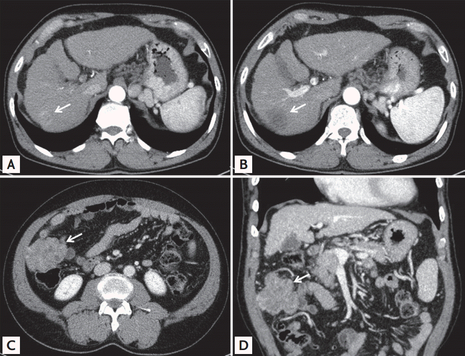
A 62-year-old male cirrhotic patient with hepatitis B was diagnosed with HCC. On the computed tomography (CT) scan, a 2-cm solitary tumor was found in hepatic segment VII, compatible with HCC (Fig. 1A). Laboratory findings showed an elevated protein induced by vitamin K absence/antagonist-II (PIVKA-II) level (586 mAU/mL), with a normal ╬▒-fetoprotein level. The patient was treated with percutaneous RFA. Artificial ascites was created for better visualization of the tumor. The treatment was successful, with complete necrosis of the tumor (Fig. 1B). Twelve months later, a 6-cm lobulated heterogeneous highly enhancing mass was found on the anterior aspect of the ascending colon on a routine follow-up CT scan (Fig. 1C and 1D). Colonofiberscopy showed no luminal mass or extrinsic compression sign.
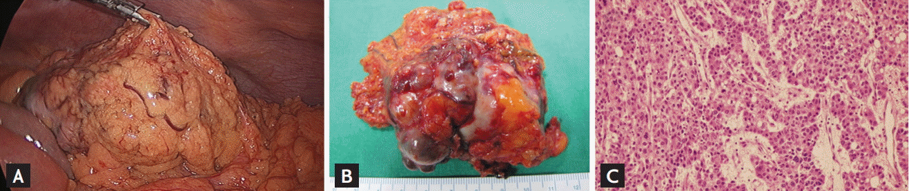
Laparoscopic exploration was performed and a 6 ├Ś 4-cm ovoid highly vascular tumor completely wrapped in the omentum was found in the right paracolic gutter (Fig. 2A and 2B). The tumor showed no colonic invasion, was easily dissected free from the surrounding tissues, and was completely removed. Pathologically, it was diagnosed as metastatic HCC (Fig. 2C). Four months after surgery, there is no evidence of tumor recurrence on CT scan, and the PIVKA-II level has normalized.
Drop metastasis after RFA may occur when handling the needle. Cancerous cells may fall and seed the peritoneal cavity forming metastatic deposits. Therefore, to prevent cancer spillage, it is important to minimize the use of artificial ascites and avoid needle entry through the free peritoneal cavity.
 PDF Links
 PubReader
 ePub Link
 Full text via DOI
 Download Citation
 Print
Print


