


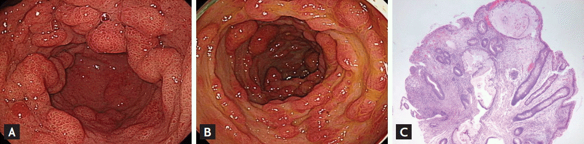
A 55-year-old man presented to our department complaining of profuse diarrhea and poor appetite for 1 month. He also reported weight loss of about 6 kg over 2 months. His medical history revealed diabetes mellitus, although well controlled. On physical examination, his fingernails and toenails were dry and dystrophic (Fig. 1A and 1B). We also noticed hyperpigmentation in both palms (Fig. 1C) and hair loss around the crown, with positive hair pull test (Fig. 1D). The laboratory studies were unremarkable, except for hypoalbuminemia (3.1 g/dL). Abdominal computed tomography showed innumerable polyps, along with reactive lymph node enlargement in the stomach, small bowel, and colon. Upper endoscopy revealed thickened mucosal folds and multiple sessile polyps of various sizes throughout the stomach and duodenum (Fig. 2A), while colonoscopy showed multiple strawberry-like polyps with nodular mucosa in the entire colon (Fig. 2B). Microscopic examinations of biopsies from these polyps revealed cystic glandular dilatation and epithelial hyperplasia; lamina propria was edematous, containing mononuclear cell infiltrates (Fig. 2C). These findings were consistent with those of hamartomatous polyps. His family history was not significant for gastrointestinal polyposis complications. Cronkhite-Canada syndrome (CCS) was diagnosed based on clinical manifestations, imaging features, endoscopic, and pathologic findings.
CCS is a very rare pathological entity. Unlike other hamartomatous polyposis syndromes, it is characterized by adult onset, non-inherited gastrointestinal polyposis, and diagnostic ectodermal findings, including alopecia, onychotrophy, and pigmentation. When encountered with a patient with innumerable polyps in the gastrointestinal tract, a clinician should have a high index of suspicion for CCS, and look for characteristic clinical, endoscopic, and histopathological findings of CCS and take special note of the different gastrointestinal polyposis conditions that may be present.

 PDF Links
 PubReader
 ePub Link
 Full text via DOI
 Download Citation
 Print
Print


