


To the Editor,
Immunoglobulin G4 (IgG4)-related disease is a recently recognized disease characterized by lymphoplasmacytic tissue infiltration by IgG4-positive plasma cells, tissue fibrosis, and elevated serum IgG4 concentration. It is usually accompanied by various clinical features such as IgG4-related autoimmune pancreatitis (AIP), sclerosing cholangitis, sclerosing sialadenitis, and retroperitoneal fibrosis [1]. But solitary tumefact tion, called IgG4-related inflammatory pseudotumor (IPT) is a rare clinical and radiological finding. Moreover, solitary IgG-related IPT of the abdomen wall is extremely rare. IgG4-related IPT is a mass-like lesion consisting of abundant infiltration of IgG4-positive plasma cells [2]. We reported a rare case of solitary IgG4-related IPT of the abdomen wall without other IgG4-related diseases.
A 54-year-old male patient who had a history of gallbladder stone in 2011 was admitted to Soonchunhyang University Seoul Hospital for epigastric pain started 1 day ago. He had no other constitutional symptoms, including fatigue, malaise, decreased appetite, weight loss, and night sweats. On admission, vital signs including body temperature were normal and physical examination showed mild right upper quadrant tenderness without definite MurphyŌĆÖs sign. On laboratory test, white blood cell count was 8,100/┬ĄL (normal range, 4,000 to 10,000), hemoglobin was 14.9 g/dL (range, 12 to 16), and platelet was 202 K/mm3 (range, 130 to 450). Aspartate aminotransferase, alanine transaminase were 34 U/L (normal range, 0 to 31), 41 U/L (normal range, 0 to 31), total bilirubin was 0.6 mg/dL (range, 0.2 to 1.2), alkaline phosphatase was 199 U/L (range, 104 to 338), ╬│-glutamyl transpeptidase was 33 U/L (range, 11 to 49), amylase was 94 U/L (range, 28 to 100), lipase was 29 U/L (range, 13 to 60), blood urea nitrogen was 9.8 mg/dL (range, 8 to 20), and creatinine was 0.90 mg/dL (range, 0.40 to 1.10). Erythrocyte sedimentation rate was 16 mm/hr (normal range, 0 to 30) and C-reactive protein was 0.27 mg/dL (normal, < 0.3). Hepatitis B and C viral antigens were negative.
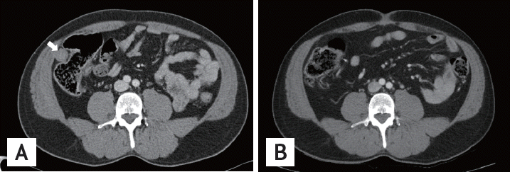
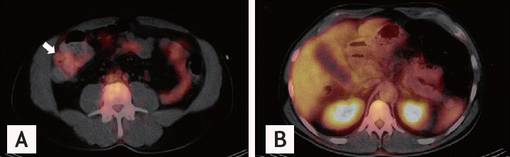
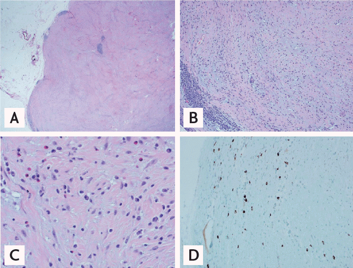
Abdomen computerized tomography (CT) showed mild distension of gallbladder with minimal pericholecystic fat infiltration which is consistent of early acute cholecystitis, but there was no other evidence of pancreatitis and sclerosing cholangitis on CT scan. Also, there was an about 2.5 cm sized subtle enhancing soft tissue density nodular lesion in right lower abdomen wall (Fig. 1A). The most common soft tissue neoplasm of the abdominal wall is the desmoid tumor. CT is the most commonly used initial imaging modality to diagnose the abdomen wall mass. For the exclusion of primary malignancy, fluorodeoxyglucose positron emission tomography (FDG-PET)/CT was done, and there was a mild FDG activity in right lower abdomen wall (Fig. 2A). Maximal standard uptake value (SUV) of the mass was 1.3. Based on SUV of the mass, we could not differentiate whether it is malignant mass or not. There was a diffuse and mild hypermetabolism around gallbladder on FDG-PET/CT which was suggesting possible cholecystitis (Fig. 2B). We operated the gallbladder and unknown origin mass in right lower abdomen wall. The histology of soft tissue in right lower abdomen wall showed fibrotic nodule with peripheral lymphoid tissue and increased plasma cells. For the differentiation between inflammatory myofibroblastic tumor (IMT) and IgG4-related IPT, immunohistochemical stains for anaplastic lymphoma kinase (ALK), IgG, and IgG4 were done. On immunohistochemical stain, IgG4-positive plasma cells were estimated up to 40/HPF and ALK was negative (Fig. 3). We checked the serum IgG4 level which was 43.5 mg/dL (normal range, 3.0 to 201). Serum IgG level was 1,156 mg/dL (normal range, 700 to 1,600), and antinuclear antibody was negative. There were no clinical features which suspect other IgG4-related organ involvement. Based on pathologic finding of the soft tissue, we diagnosed solitary IgG4-related IPT of the abdomen wall. After 4 months, we checked abdomen CT scan for follow-up, there was no newly developed mass-like lesion in the abdomen and pelvis (Fig. 1B).
The incidence of IgG4-related disease throughout Japan was estimated to be 0.28 to 1.08/100,000, with 336 to 1,300 patients newly diagnosed per year and approximately 6,700 to 26,000 patients who developed IgG4-related disease over the past 20 years. It predominantly affects middle-aged to elderly men. As IgG4-related disease affects various organs, it presents variety of clinical symptoms. Patients often feel well at the time of diagnosis and generally lack fever. However, patients with multiorgan disease often lose weights (about 9 to 14 kg) over several months. Many of the initial symptoms regarding this condition were made in patients with AIP, which often presents as a pancreatic mass or as painless obstructive jaundice. Other patients with sclerosing sialadenitis may present with prominent parotid or submandibular gland enlargement [1]. Asymptomatic IgG4-related lymphade-l nopathy is also common. It is usually observed together with other clinical manifestations of the syndrome, but may be the initial or only manifestation [3].
The diagnosis of IgG4-related disease is based upon biopsy findings demonstrating the characteristic histopathologic findings and immunohistochemical staining. Diagnostic criteria are established with two major features of IgG4-related disease: elevated serum level of IgG4 and infiltration of IgG4-positive cells. The minimum value for serum IgG4 level is 135 mg/dL. Histopathological features are IgG4-positive plasma cell infiltration (> 10 cells/HPF) and/or IgG4-positive/IgG-positive cell ratio > 40%. In order to diagnose the IgG4-related disease, it is necessary to biopsy the suspicious lesion [1]. Although serum IgG4 level is included in the diagnostic criteria, about 3% to 30% of IgG4-related disease patients have normal serum IgG4 concentrations. Thus the histopathologic findings of the lesion are essential to diagnose the IgG4-related disease.
IgG4-related IPT can occur in various organs such as pancreas, liver, lung, heart, kidney, orbit, gastrointestinal tract, but its incidence is relatively lower than systemic manifestation of IgG4-related diseases [4]. Solitary IgG4-related IPT without lymphadenopathy can mimic malignancies, it is important to differentiate this lesion with other type of mass-like diseases. IMT is a rare neoplasm of intermediate biologic potential that usually arise in children to young adults. IPT and IMT share some morphological features including prominent fibroblastic/myofibroblastic proliferation and the presence of inflammatory cells. Since IgG4-related IPT and IMT require different treatment modalities, it is necessary to differentiate these two lesions. IgG4-related IPT is treated conservatively with corticosteroid, whereas IMT should undergo surgery due to its malignant potency. Approximately half of IMTs carry rearrangements of the ALK causing aberrant ALK expression [5].
Our case was a mass like lesion in the abdomen wall which was identified with acute cholecystitis accidentally. We underwent surgery and the center of the mass was fibrotic and contained fibroblasts and a few infiltrated inflammatory cells including plasma cells, lymphocytes and eosinophils. Immunohistochemically, most plasma cells were IgG4-positive and the mass showed negative finding for ALK. We diagnosed the solitary IgG4-related IPT and removed the abdomen lesion surgically. Four months after the surgery, patient had no newly developed lesion and no others symptoms which were suspicious of IgG4-related disease without medical treatment including corticosteroids.
 PDF Links
 PubReader
 ePub Link
 Full text via DOI
 Download Citation
 Print
Print


