


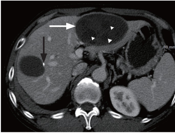
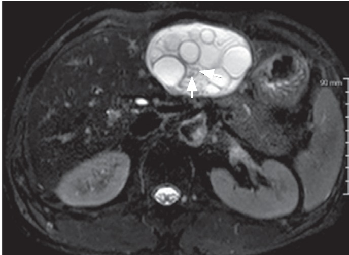
A 58-year-old man was admitted for the evaluation of an incidentally detected hepatic mass. Intravenous contrast-enhanced abdominal computed tomography scan in conjunction with contrast-enhanced T1-weighted magnetic resonance imaging (MRI) showed a smooth-margined cyst in the left lateral hepatic segment, which measured 8.5 cm in diameter at the widest point, and contained several other small cysts. There was also another sizable cyst in right hepatic lobe, measuring roughly 3.3 cm in diameter, with a small calcification along the wall (Fig. 1). T2-weighted MRI showed additional small low signal intensity tubular lesions within the large cyst (Fig. 2).
At the time, the patient's laboratory findings, including white blood cell count, differential eosinophil count, C-reactive protein, and liver function test, were within normal range. He often ate canine flesh and pork; however, his stool examination was negative for parasite egg.
The patient underwent left lateral hepatic sectionectomy and right hepatic tumorectomy. The gross morphology of the large cyst in the left lateral hepatic segment showed fibrotic material, several small gelatinous fluid collections, serous fluid, and a few small dead tubular worms. They were identified pathologically as Echinococcus granulosus. The other cyst in right hepatic lobe was also considered as a hepatic hydatid cyst.
Echinococcosis in humans is a zoonotic secondary infection by the larval stages of the cestode species of the genus Echinococcus. Cystic echinococcosis (CE) is caused by E. granulosus. The intermediate host, humans, enter the organism's life cycle through exposure to infected canine feces. If the cyst contains a living worm, peripheral eosinophilia can be frequently seen. The liver is the most common organ of CE. In the case of hepatic CE, the right hepatic lobe is more frequently involved, and the morphological form, about 75% of cases, is a unilocular simple cyst.
However, a large mother cyst containing multiple small daughter cysts in the liver is considered relatively typical and highly specific imaging finding for the diagnosis of hepatic hydatid cyst. In this case, there were a few small tubular structures within the cyst on MRI were regarded as dead worms in the pathologic specimen. A second cyst with a small calcification is non-specific imaging finding. In the presented case, it was another hepatic hydatid cyst. According to the classification of a hepatic hydatid cyst, a cyst with calcification is considered a dead cyst.
 Print
Print


