


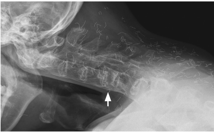
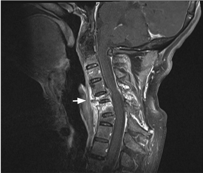
A 51-year-old man with a 22-year history of ankylosing spondylitis who had been treated with nonsteroidal anti-inflammatory drugs and infliximab presented with severe posterior neck pain, which had been aggravated after he fell backwards 3 months earlier. The neurological examination was normal. Plain radiographs showed total ankylosis of the cervical spine and a C5 vertebral body fracture with anterior dislocation of C4 on C5 (Fig. 1). There were numerous thin pieces of foreign material along the spine (Fig. 1). The patient said he had undergone Korean traditional gold thread acupuncture therapy, which had not been effective. Gadolinium-enhanced fat-suppressed T1-weighted magnetic resonance imaging showed a recent compression fracture of the C5 vertebral body and resultant spinal cord compression (Fig. 2), as well as an old compression fracture of C7. For cervical spine fractures in ankylosing spondylitis, conservative treatment with gentle low-weight cervical traction can be performed initially, in the absence of a neurologic deficit. However, secondary neurological deterioration occurs frequently due to the delayed dislocation at the original fracture site, and surgical treatment is generally recommended. On surgical exploration of our patient, the C5 fracture was found to be stable and surgical f ixation was not done. The cervical spine is the most frequent site of acute spinal fracture in ankylosing spondylitis, particularly at C5-6 and C6-7. Since the cervical fractures in ankylosing spondylitis are usually unstable, with a high risk of neurological deficits, our patient was lucky not to have had any significant neurological complications.
 PDF Links
 PubReader
 ePub Link
 Full text via DOI
 Download Citation
 Print
Print


 |
 |