


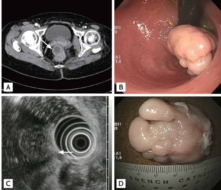
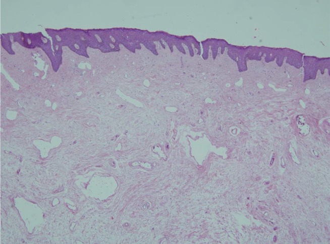
A 48-year-old woman was referred to our hospital for evaluation of a large subepithelial rectal mass, which had been incidentally detected on abdominopelvic computed tomography (CT) (Fig. 1A). Two years prior, she had undergone hepatopancreaticoduodenectomy with right hemicolectomy to treat gallbladder cancer that had invaded the liver and ascending colon. Subsequently, she received adjuvant chemotherapy and annual follow-up. CT revealed a 3-cm-diameter large pedunculated subepithelial mass covered by intact whitish mucosa in the distal rectum (Fig. 1B). On endoscopic ultrasonography (EUS), a heterogenously hypoechoic lesion with scattered hyperechogenic spots was observed in the deep mucosal and submucosal layers (Fig. 1C). The CT and EUS findings raised the suspicion of an inflammatory fibrinoid polyp, a lymphangioma, or a rare form of metastatic cancer. She underwent endoscopic mucosal resection using a detachable snare. The resected specimen was approximately 4 cm in diameter and had a nodular shape (Fig. 1D). Pathologically, the tumor consisted of a fibrous stroma with small dilated vessels, and was covered with normal squamous epithelium (Fig. 2). The final diagnosis was a rectal fibroepithelial polyp. Recurrence was not observed on follow-up colonoscopy performed 2 years later.
Fibroepithelial polyps are common benign polypoid tumors formed by squamous epithelium and subepithelial connective tissue. They are generally small in size and asymptomatic. However, a giant fibroepithelial polyp can cause intussusception or obstructive ileus. Therefore, if a tumor is larger than 3 cm in diameter, endoscopic or surgical resection is recommended. Follow-up is not essential, because no case of malignant transformation of a fibroepithelial polyp is known.
 PDF Links
 PubReader
 ePub Link
 Full text via DOI
 Download Citation
 Print
Print


