


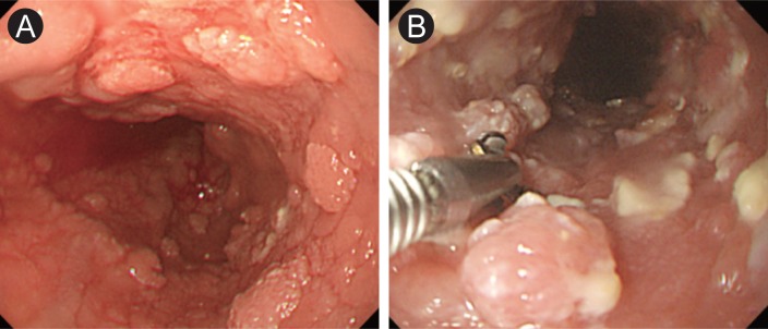
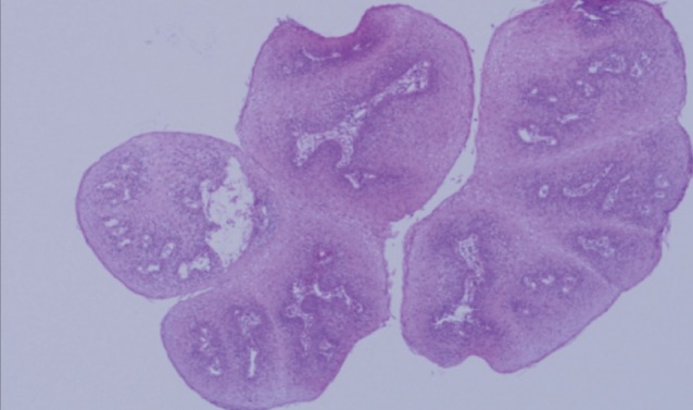
A 69-year-old man underwent upper endoscopy at our hospital to evaluate gastric discomfort. He had undergone a subtotal gastrectomy for advanced gastric cancer 2 years earlier, but had been lost to follow-up. An endoscopic examination revealed numerous (> 100) sessile to polypoid mucosal nodularities. Their mucosa had a characteristic verrucous appearance similar to papillomatous skin warts extending from the piriform sinus to the esophago-gastric junction (Fig. 1). Their size ranged from 1 to 10 mm. Histologically, biopsies taken at multiple levels revealed papillary projection lined with acanthotic squamous epithelium. This finding was consistent with squamous papilloma without evidence of dysplasia or carcinoma (Fig. 2). A human papilloma virus (HPV) test using a HPV DNA chip kit was negative. He was transferred to another hospital and was shortly lost to follow-up.
Esophageal squamous papilloma (ESP) is a rare benign epithelial lesion characterized histologically by finger-like projections of hyperplastic squamous epithelium covering a connective tissue core. Most lesions are small, solitary, and found incidentally. It is rarely difficult for an experienced endoscopist to identify ESP because of the typical warty appearance. ESP is extremely rare and has been described in fewer than 10 cases in the English-language literature. The etiology of ESP remains unclear, although roles of chronic mucosal irritation and HPV infection have been proposed. The clinical course of ESP is variable, ranging from natural regression to the development of squamous cell carcinoma. A slowly progressive course appears to be most common. Therefore, clinicians should be aware of the malignant potential of these lesions. However, there are no standard therapeutic or surveillance guidelines for esophageal papillomatosis due to the scarcity of reported cases.
 PDF Links
 PubReader
 ePub Link
 Full text via DOI
 Download Citation
 Print
Print


