INTRODUCTION
Intrahepatic stones occur commonly in East Asia, and intrahepatic stones retained or recurred after surgery represent a major problem1). Retained or recurred stones after surgery can be removed by mechanical manipulation, either with fluoroscopic guidance or by direct vision with the flexible cholangioscope. But the results have not been always satisfactory.
Extracorporeal shockwave lithothripsy (ESWL) has been applied to intrahepatic stones. However, the effectiveness and safety of this technique requires confirmation due to small patient series in most of the reports2–7). We have studied the efficacy of ESWL in 18 patients with multiple intrahepatic stones.
MATERIALS AND METHODS
During the period of twenty months from July 1989 to February 1991, we have treated 18 patients of intrahepatic stones with ESWL. The locations of stones were right intrahepatic bile ducts in 2 patients and bilateral intraheptic bile ducts in 16 patients. Their stone numbers were more than 10, and all patients had strictures of the biliary tract. All patients (11 male, 7 female, average age 45 years) received operations previously due to intrahepatic stones.
Using the dual ultrasound system of the Dornier Lithotripter MPL 9,000, intrahepatic stones were localized and positioned in the shockwave focus. Discharge voltage was average 17 KV. All patients were treated in a prone position and general anesthesia was not given. When necessary, 50 mg pethidine was given intramuscularly.
The patients with intrahepatic stones had T-tube (n = 9) or percutaneous transhepatic biliary drainage (PTBD) tube (n = 9). Patients with PTBD or T-tube received balloon dilatation of stricture after complete stone removal.
RESULTS
In all cases, the intrahepatic calculi were successfully localized and positioned in the shockwave focus. Each patient received an average of 4 treatments, resulting in a total number of 3,064–12,000 shockwaves per patient (average 6,288 shocks).
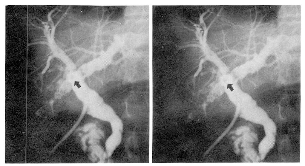
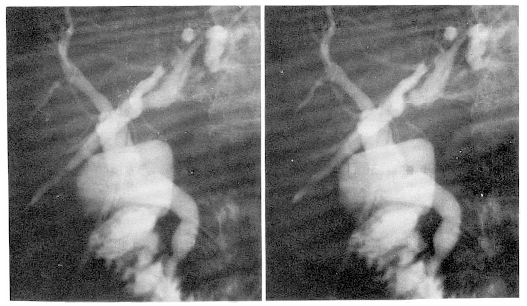
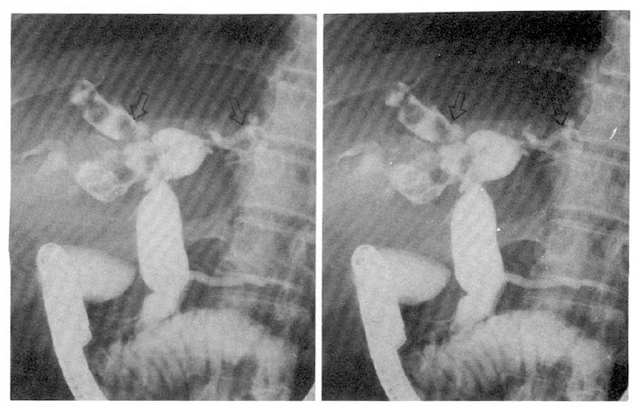
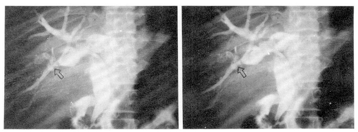
Intrahepatic stones were successfully disintegrated in all patients and the disintegrated stones were removed successfully in sixteen patients by subsequent cholangioscopic or interventional radiologic stone extraction. Among nine patients with indwelling T-tube in the common bile duct, four patients became completely stone free, with ESWL therapy only. After ESWL treatment, fragments passed spontaneously through strictures of the intrahepatic biliary tree. Then multiple stone fragments and sludge passed through the T-tube. Three additional patients became stone free after ESWL and cholangioscopic extraction of debris from the biliary system. But in two patients, complete stone removal was impossible and partial reduction of intrahepatic stone volume was achieved.
In the nine patients with PTBD, all patients became stone free after combination of ESWL and basket extraction via PTBD track. The large impacted stone that had previously obstructed the intrahepatic duct just above the stricture was easily dislodged by ESWL and the majority of more proximally located remaining stones could be successfully manipulated by basket.
Nine patients had mild transient hemobilia during ESWL treatment. In 2 cases, temporary sepsis developed. But no other significant complications were observed.
DISCUSSION
Primary hepatolithiasis or oriental cholangiolithiasis is common in South-East Asia and remains a very difficult problem to manage. Retained or recurred stones in the intrahepatic ducts after surgery are frequent and troublesome7,8). Bilateral intrahepatic biliary stones with multiple strictures have ofter proved unbeatable in the past. Postoperative cholangioscopy via T-tube sinus tract and percutaneous transhepatic intervention with fluoroscopic guidance have been the useful alternative means for treating hepatolithiasis9–12). However, there are many cases in which the complete removal of intrahepatic biliary stones cannot be accomplished, even by cholangioscopic or percutaneous interventional approach. There have been two problems leading to such unsuccessful results in the treatment with cholangioscope via T-tube sinus tract. The first is how to insert the endoscope through the biliary stricture and reach the impacted stones. The second is how to fragment the stone in the unfavorable location. Sharp angles and severe strictures of the biliary ducts containing stones result in treatment failure13). Our observation that fragmented intrahepatic stone (in front of stenoses) by ESWL passed spontaneously through strictures and drained via T-tube was surprisingas it was unexpected. If further therapy of this disease confirms these observations, then ESWL might become a new treatment modality for oriental cholangiolithiasis.
In the percutaneous radiologic intervention with stone basket via PTBD track, it is difficult or impossible to manipulate the impacted stone proximal to stricture. In our patients with PTBD, stone grasping by basket via PTBD track was not successful in the case of impacted stone just above the stricture. After ESWL with focusing on impacted stone in front of stenosis, it became easy to grasp and fragment the impacted stone by basket and manipulation of the stones more proximally located has become possible. Surprisingly, impacted stone, which was initially observed to be unaffected by the shocks, disintergrated after being touched and manipulated with the stone basket. Sometimes, we have observed that ESWL of intrahepatic stone just above the stricture made it possible for the fragmented debris to pass through the stricture spontaneously. Any procedure suitable for the treatment of intrahepatic lithiasis must include both complete removal of stones and establishment of adequate biliary drainage to relieve bile stasis17). Our patients with PTBD or T-tube received balloon dilatation of stricture after stone removal.
Our results show that ESWL is very effective in facilitating extraction of stones in unfavorable locations or located in front of severe stricture. Although complete freedom of stones can be reached after multiple sessions, ESWL followed by percutaneous stone extraction will provide an improvement in the success rate and duration of treatment required for complete removal of intrahepatic stones.


 PDF Links
 PubReader
 ePub Link
 Full text via DOI
 Download Citation
 Print
Print


