


INTRODUCTION
Left ventricular diastolic function is a more sensitive indicator of left ventricular dysfunction than the systolic one in coronary artery disease,1–3) and can be evaluated by several noninvasive methods such as radionuclide ventriculography3,4) or M-mode echocardiography.5)
Doppler echocardiography, widely applied in the hemodynamic evaluation of various cardiac diseases, has been used to assess the left ventricular diastolic performance using the transmitral flow velocity curve, which has been reported to reflect left ventrivular filling characteristics relatively well compared with contrast6) or radionuclide ventriculography.7) However, it is not certain that pulsed Doppler echocardiography is so sensitive to detect disturbed diastolic function even in patients with angina pectoris who have no evidence of left ventricular hypertrophy or abnormal systolic function, although there have been several reports revealing that pulsed Doppler echocardiography can assess the altered left ventricular diastolic filling in patients with cardiomyopathy,8,9) hypertension,10) or coronary artery disease.11–13)
The purpose of this study was to evaluate whether pulsed Doppler echocardiography could be useful in the detection of disturbed diastolic function in patients with angina pectoris who have normal systolic function and no left ventricular hypertrophy by analysis of the transmitral flow velocity curve.
MATERIALS AND METHODS
1. Study Population
Of all the consecutive patients who received both coronary arteriography and echocardiographic examination due to anginal or atypical chest pain between February and October 1986 at Seoul National University Hospital, 55 subjects, who met the following criteria were prospectively selected as a study population. Criteria for inclusion in this study were 1) over 40 years of age, 2) no evidence of left ventricular hypertrophy on M-mode echocardiogram (thickness of interventricular septum and left ventricular posterior wall<13 mm), 3) normal regional wall motion on contrast left ventriculography, and 4) no other associated cardiovascular disease such as valvular, myocardial, or pericardial disease. Patients with moderate to severe hypertension or a history of myocardial infarction were also excluded.
The study population was devided into two groups, the angina and control groups. The angina group, which coronary arteriography showed significant narrowing (⩾ 75% of luminal diameter) in one or more of the major coronary arteries. The extent of disease was one vessel disease in 16, two vessels in 9, and three vessels in 8 patients. The control group, which comprised 22 subjects, was defined as a group of subjects who had no (n = 18) or insignificant narrowing (<50% of luminal diameter) (n = 4) on coronary arteriography.
Each subject was not restricted from medication such as nitrates, calcium antagonists, or beta blockers and was in normal sinus rhythm at the time of both procedures.
2. Angiographic Methods
All the cineangiographic studies were performed with the use of the percutaneous femoral technique, frame rates of 60 frame/sec, and were analyzed by at least two experienced cardiologists who were unaware of the echocardiographic results. Single plane left ventriculography was done before coronary arteriography with the patients in the 30 degree right anterior oblique position after the injection of 40 to 45 ml (10 to 12 ml/sec) of contrast media (Telebrix 38). A 1 cm grid was filmed for the correction of magnification. The regional wall motion of the left ventriculogram was analyzed with the use of a cineangiographic projector (Tagarno 35D) and the ejection fraction of the left ventricle was calculated with the use of the area-length method by Sandler and Dodge.14)
Coronary arteriography was done with Judkins’ method in the multiple projections and the degree of the lesion was determined in % narrowing of luminal diameter of coronary artery.
3. Echocardiographic Studies
Echocardiographic examination was performed one day before angiographic study in each patient with a stable clinical condition. Commercially available Aloka SSD-880 CW system (Aloka Inst., Japan), which has M-mode, two dimensional (90 degree) phased array sector scanner with a pulsed Doppler flow transducer of 2.5 MHz, was used for the recording of transmitral flow velocity. The examination was done with the patient in the left lateral position and all recordings were made at the end of expiration with the paper speed of 100 mm/sec using stripchart recorder (SSZ-95, Aloka Inst., Japan).
Transmitral flow velocity curve by pulsed Doppler echocardiography was obtained with an apical four chamber view which provided good visualization of left ventricular cavity and mitral valve leaflets. The sample volume was located at the tip of the mitral valve leaflet, slightly below the mitral annulus, where a maximal Doppler flow velocity signal of good quality can be obtained, and care was also taken to minimize the intercept angle between the directions of the Doppler beam and presumed transmitral flow.
4. Quantitative Analysis of the Transmitral FlowVelocity Curve
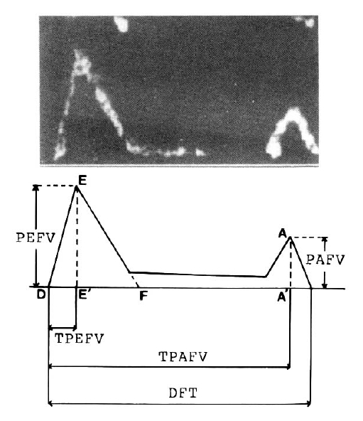
In each patient diastolic transmitral flow velocity curves of at least three cardiac cycles were analyzed and averaged. The following parameters of transmitral flow velocity were measured as shown in figure 1. 1) Peak flow velocity during early diastole (E-E’, peak early flow velocity, PEFV), peak flow velocity during late diastole by atrial contraction (A-A’, peak atrial flow velocity, PAFV), and the ratio of early to late peak flow velocities (E-E’/A-A’, PEFV/PAFV); 2) The descending slope of peak early flow velocity (EF slope in cm/sec2) is the rate of decrease in early diastolic flow velocity after peak; 3) Time intervals from the onset of diastolic flow to the peak early flow velocity (D-E, time to peak early flow velocity, TPEFV), to the peak atrial flow velocity (D-A, time to peak atrial flow velocity. TPAFV), and to the end of diastolic flow (total diastolic filling time, DFT).
5. Reproducibility
Interobserver and intraobserver reproducibility of parameters of transmitral flow velocity curve were evaluated in 22 study subjects (12 without and 10 with significant coronary artery stenosis). Measurements from three cardiac cycles were averaged and compared. Correlations were excellent for various parameters measured by the same observer on separate occasions (r>0.97 for each variable) and by two different observers (r>0.95 for each variable).
RESULTS
There were no statistically significant differences between the control and angina groups in the various clinical characteristics including age, sex, mean heart rate, blood pressure, cardiac index, angiographic ejection fraction and left ventricular end diastolic pressure (Table 1).
Transmitral Flow Velocity Curve
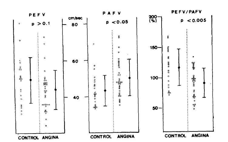
Diastolic parameters derived from the transmitral low velocity curve of the two groups were compared (Table 2). In the angina group, peak atrial flow velocity (50.1 ± 10.0 cm/sec) was higher than that of the control group (43.7 ± 9.0 cm/sec) (p<0.05), but there was no significant difference between the peak early flow velocities of both groups. The ratio of early to atrial peak flow velocity in the angina group (0.91 ± 0.24) was lower than that of the control group (1.17 ± 0.30) (p<0.005) (Figure 2). Other parameters including normalized time to peak early or atrial flow velocity and deceleration slope of peak early flow velocity (EF slope) were not significantly different between the two groups.
DISCUSSION
Left ventricular diastolic filling has been reported to be impaired in many patients with angina pectoris, even though they have no evidence of systolic dysfunction.1–4) The mechansm of disturbed diastolic function is not clear, but incomplete or impaired relaxation,15) altered diastolic tone,16) tension prolongation during recovery from hypoxia,17) and asynchronous left ventricular diastolic filling4) have been suggested as possible mechanisms. Among the many indices of left ventricular diastolic function, peak filling rate during early diastole has been widely used to assess the disturbed left ventricular diastolic filling in patients with coronary artery disease, because it cannot only be obtained easily by noninvasive methods but reflects left ventricular relaxation relatively well.3,5,18) Recently, Doppler derived the peak transmitral flow velocity has been proposed as an useful index of left ventricular diastolic function in patients with several heart diseases, but little is known about its ability to detect disturbed diastolic filling sensitively in angina patients with normal systolic function.
The pulsed Doppler echocardiographic technique of evaluating the left ventricular diastolic function has several advantages despite a few limitations such as poor echocardiographic window, spectral widening, and intercept angle between directions of Doppler beam and transmitral flow, etc. Major advantages of the Doppler method of analyzing the transmitral flow velocity curve are that transmitral flow velocity is not only independent of left ventricular size or geometry19) but also able to assess left ventricular filling on a beat to beat basis with excellent temporal resolution.
Of the several parameters derived from the transmitral flow velocity curve, the ratio of early to atrial flow velocity is thought to be most sensitive,7,12) although peak atrial flow velocity alone is also useful. Recently, the integrated area under the transmitral flow velocity curve during the first one third or half of the diastolic filling period has been suggested as another useful index of rapid diastolic filling,8,13) but it seems to have little advantage over the previous one because of errors associated with calculation or digitization of the integrated area.
Our results of a lower ratio of early to atrial flow velocity, and higher peak atrial flow velocity in the angina group were similar to those of Wind et al.13) But we couldn’t find significant difference in peak early flow velocity between both groups, whereas Wind et al. reported a lower peak early flow velocity in patients with coronary artery disease. Such a difference seems to be due to the difference of study subjects between the two studies, that is, patients with prior myocardial infarction or wall motion abnormalities were included in the study by Wind et al., but excluded in this study. The above results may be used to say that the pulsed Doppler echocardiographic method of analyzing the transmitral flow velocity curve is very useful, and the ratio of early to atrial peak flow velocity and peak atrial flow velocity is more sensitive index than peak early flow velocity in the evaluation of left ventricular diastolic function.
However, values of peak atrial flow velocity or the ratio of early to atrial flow velocity showed an overlap of considerable degree in spite of a statistically significant difference between the two groups. A few possible factors which can influence the diastolic function must be considered in interpretating this data as well as others which assess the diastolic function.
Firstly, the left ventricular diastolic function, is impaired in many but not all angina patients with normal systolic function, even in the presence of significant coronary arterial narrowing, and the range of normal diastolic function is so wide that an overlap of considerable degree is inevitable between the two groups, especially when examined at rest. Similar results have been reported by other studies using radionuclide ventriculography.3,4)
Secondly, drugs, especially antianginal drugs such as nitrates or calcium antagonists, can be a possible factor of overlapping. In this study, the use of antianginal medication was not limited at the time of Doppler echocardiography or coronary arteriography but this factor can’t explain the difference of diastolic parameters between both groups, because nitrates or calcium antagonists are known to improve the diastolic function in patients with coronary artery disease.
Thirdly, the difference in the extent of left ventricular wall thickness or size could be a source of overlap or difference in the results between both groups, because wall thickness, size or the ratio of wall thickness to chamber size are known to influence the chamber stiffness,20) which is one of the major determinants of left ventricular filling. However, this isn’t likely to explain the difference, because we couldn’t find any statistical difference in thickness of the interventricular septum or left ventricular posterior wall and we also excluded patients who had a dilated or abnormally hypertrophied left ventricle by two dimensional echocardiography or contrast ventriculography.
The age factor also isn’t likely to explain the difference in the Doppler diastolic parameters because the two groups of patients were similar in age, although age has been reported to affect the transmitral flow velocity.21)
In conclusion, the pulsed Doppler echocardiographic technique analyzing the transmitral flow velocity curve seemed to be useful for noninvasive assessment of the disturbed left ventricular diastolic filling in patients with angina pectoris who have normal systolic function.
 PDF Links
 PubReader
 ePub Link
 Full text via DOI
 Download Citation
 Print
Print


