Korean J Intern Med > Volume 2(1); 1987 > Article
Kim, Heo, Bang, Lee, Choi, Park, Kim, Kim, Heo, and Lee: Multiple Myeloma in Korea*

Abstract

A clinical analysis was made of 61 cases of multiple myeloma diagnosed between January 1976 and June 1984 at Seoul National University Hospital using the criteria of the Southwest Oncology Group.
The following observations were made.
  • 1) The major clinical problems at Initial presentation were bone pain(52%), anemia(20 %), renal impairment(11%), and infection(10%).

  • 2) Clinical stages at presentation were stage I, 11%; stage II, 8%; and stage III, 81% of patients. Three(5%) of the patients in stage II and 18(31%) in stage III showed renal impairment with a serum creatinine ≥2.0 mg/dl.

  • 3) Combination chemotherapy produced a response rate of 29% with melphalan and prednisone, and 40% with M2 protocol(among 14 and 20 evaluable patients respectively). Both regimens showed statistically significant survival difference between responders and non-responders(p <0.01).

  • 4) The median survival of all patients was 13 months.

  • 5)Age, calcium level, creatinine level, and performance status were important prognostic factors on survival.

INTRODUCTION

Multiple myeloma is one of the most common plasma cell neoplasms and mainly involves bone and bone marrow. It is not an uncommon disease in Korea. Previous studies in Korea included only small numbers of cases. In 1972 an initial statistical analysis of 28 cases was presented at the Korean Hematology Meeting1). A second report was made at the 4th International Hematology Meeting of Asia and Pacific Area2). Additional studies concentrating on the M-protein were reported at the Korean Hematology Meeting in 19833). As of this writing, there has been no Korean report on the results of chemotherapy.
We, therefore, analyzed the clinical features and chemotherapeutic results of 61 patients with multiple myeloma who were diagnosed between January 1976 and June 1984 at Seoul National University Hospital.

MATERIALS AND METHODS

Between January 1976 and June 1984, 61 consecutive patients were diagnosed as having multiple myeloma at Seoul National University Hospital. The diagnosis was established according to the criteria of the Southwest Oncology Group (SWOG)4). The patients were clincally staged using the system developed by Durie and Salmon5).
Twenty one patients were treated with melphalan and prednisone (MP, Table 1), and 25 patients with the M2 protocol (Table 2)6). An objective response was defined as a reduction of 50% or more in serum M-protein concentration or in urine 24 hour light chain excretion7). This protein response must be accompanied by normal serum calcium, serum albumin above 3 g/dl, and no progression of skeletal disease. Determination of treatment effect was limited to patients who had received at least 2 cycles of chemotherapy, and whose M-protein levels had been continuously monitored. Remission duration was defined as the period from the day when the M-protein concentration decreased to less than 50% of the pretreatment value to the day when it doubled from the lowest value obtained during the remission period. Survival curves were calculated from the start of the therapy using the Kaplan-Meier product limit method, and the log-rank test was used for the comparison of survival curves.

RESULTS

1. Clinical Features

Patient ages ranged from 15 to 81 years (median age: 54 years). Fifty five (90%) of the 61 patients were 40 years or older. The male to female ratio was 2.8: 1 (Table 3).
Bone pain (52%) and anemia (20%) were the most common clinical problems at the time of initial presentation. Eleven percent of the patients presented with renal problems including acute or chronic renal failure and 10% with infection manifested by pneumonia, urinary tract infection or sepsis (Table 4).
Twelve cases (20%) showed plasmacytomas on biopsy of bone or soft tissue. Thirty nine cases (65%) showed bone marrow plasmacytosis which occupied more than 10% of all the nucleated cells.
Protein electrophoresis and Immunoelectrophoresis of serum and urine was done in 55 patients, and the M-protein spike was demonstrated in 50 patients. IgG was the most common type accounting for 25 cases (50%), IgA for 14 cases (28%), IgD for 4 cases (8%), and light chain only for 7 cases (14%). The ratio of kappa light chain to lambda light chain was 1.1:1. In 15 patients (25%), concentration of normal immunoglobulins was reduced. Three patients presented as solitary plasmacytoma of bone, 1 as an extramedullar plasmacytoma of the maxillary sinus, and 1 as a nonsecretory myeloma.
Forty one (68%) of the 61 patients showed osteolytic bone lesions, and 4 (6%) showed osteoporosis only on skeletal survey. In 16 patients (26%) the bone X-ray revealed no abnormalities (Table 5).
Nineteen patients (31%) had hemoglobin values over 10 g/dl, and 25 (41%) below 8.5 g/dl. Nine (15%) showed serum calcium levels over 13.0 mg/dl, and 4 (6%) between 11.0 mg/dl and 13.0 ma/dl. Those with serum creatinine level equal to or greater than 2.0 mg/dl were 21 (34%), and those below 1.4 mg/dl were 32 (52%).
Seven patients (11%) were classified as stage 1, 5 (8%) as stage II, and 49 (81%) as stage III. Three (5%) of the patients in stage II and 18 (30%) in stage III showed renal impairment with a serum creatinine ≥2.0 mg/dl (Table 6).

2. Results of Treatment

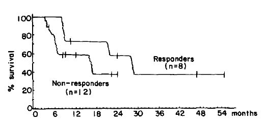
Out of 21 patients who were treated with MP therapy, 14 were evaluable. Among them, 4 patients (29%) achieved objective responses, and the median duration of response was 6 months. Twenty out of 24 patients who received the M2 protocol were evaluable. Objective responses were obtained in 8 patients (40%), and the median duration of response was 20 months. Chemotherapy related toxicity was not uncommon, but usually self-limiting and no treatment-related deaths were observed (Table 7). The median survival of the overall patients was 13 months (Fig. 1). There were statistically significant survival differences between responders and nonresponders to MP therapy (30 months vs 8 months, p<0.001, Fig. 2), and to M2 protocol treatment (28 months vs 16 months, p<0.01, Fig. 3).
Age, calcium level, creatinine level and performance status were important prognostic factors on survival. Patients with IgA M-protein survived longer than those with light chain only or IgG M-protein (Table 8).
Thirteen patients died at our hospital. The causes of death were as follows; acute renal failure with pulmonary edema in 6, sepsis in 4, intractable bleeding in 2, and airway obstruciton in 1.

DISCUSSION

The annual incidence of multiple myeloma in the U.S.A. is 3 per 100,000 population, with a peak occurrence at ages 50 to 70 year8). This disease rarely occurrs before 40 years of age, and is slightly more common in males than in females. The incidence in Korea is unknown, but it comprises about 0.3% of all malignancies in Korea9). In this study, the peak incidence was at ages 50 to 59 years with a median of 54 years, and only 6 patients (10%) were younger than 40 years of age. The male to female ratio was 2.8 : 1.
Patients with myeloma have variable clinical manifestations. Bone pain is present in 68% of patients at diagnosis, anemia in 62%, and renal problems in 50%10). Renal problems include proteinuria, tubular dysfunction, urinary tract infection, acute renal failure and chronic renal failure. Infection is frequent due to an abnormal immune system. The overall incidence is 1.46 infectious episodes per patient-year, and nearly 70% of patients with myeloma die as a result of infection11). The two months immediately following the start of initial chemotherapy was the most hazardous period. In this study, the most common clinical problems at initial presentation were bone pain (52%) and anemia (20%). Other problems include acute or chronic renal failure (11%), and infection (10%). Renal failure and infection were the most common causes of death.
Kyle et al. reported the relative distribution of immunoglobulin classes by serum and/or urine immunoelectrophoresis10). It was IgG 59%, IgA 23%, IgD 1%, and light chain only 17%. In this study, it was IgG 50%, IgA 28%, IgD 8%, and light chain only 14%.
There are many prognostic factors influencing patient survival, among which serum creatinine level is considered the most important. Other prognostic factors include performance status, hemoglobin level, serum albumin and calcium level, degree of osteolytic lesion, and amount of M-protein. The clinical staging system was developed on these parameters5). Santoro et al. reported that the median survival from diagnosis was 48 months for stage I, 41 months for stage II and 23 months for stage III12). The median survival of patients with normal renal function was 35 months compared to 7 months for those with renal impairment. Response to chemotherapy is another important prognostic factor on patient survival12,13). Patients who obtained objective responses survived much longer than those who did not. In this study, age, calcium level, creatinine level, performance status, and response to chemotherapy were found to be important prognostic factors. The median survival of patients with myeloma has been reported as 2 to 3 years8). In this study the median survival of patients was 13 months. This poor result might be explained by the fact that the majority of patients in this study were in advanced stages at the initial presentation.
Treatment of multiple myeloma is composed of general supportive care and chemotherapy. Supportive care aims to preserve ambulation and mobility, control pain and avoid dehydration. Management of complications, i.e., control of hypercalcemia, skeletal destruction, infection and renal dysfunction is also an important aspect of supportive measures. However, definitive treatment of myeloma calls for chemotherapy. Current drugs shown to be effective as a single agent include alkylating agents (melphalan, cyclophosphamide, chlorambucil), nitrosoureas, procarbazine, and doxorubicin, each of which has a remission rate of about 20–50%1416). To improve the remission rate and to prolong the survival time many combination chemotherpay regimens were investigated. One of the most commonly employed regimens is a combination of melphalan and prednisone (MP), which showed a remission rate of 48% and remission duration of 24 months13). The best reported results of combination chemotherapy are those based upon the M2 protocol developed by investigators at Memorial Sloan Kettering Cancer Center6). This regimen combines melphalan, prednisone, cyclophosphamide, vincristine, and BCNU. A remission rate of 78–87% and median survival of 38–50 months were reported6,17). The Eastern Cooperative Oncology Group compared this M2 protocol with MP therapy in a randomized study. Objective responses were seen in 74% of the patients receiving the M2 regimen, and in 53% of those receiving the MP regimen. But the median survival did not differ at 33 and 29 months, respectively18). In our study, the MP regimen produced a 29% remission rate, and the M2 protocol 40%. This lower remission rate could be explained by the large numbers of patients in advanced disease in our stydy. When patients fail to respond to initial therapy or relapse after responding to first line drugs, second-line treatment may be tried. A combination chemotherapy with BCNU and doxorubicin was shown to have a response rate of 54% in relapsed patients19). We treated 5 patients, 4 initial nonresponders and 1 relapsed patient, with BCNU and doxorubicin, but none of them responded. We think that more systematic and continous studies are required on this subject in Korea.

Fig. 1.
Survival curve of all patients.
kjim-2-1-120-20f1.gif
Fig. 2.
Survival curves by response to MP regimen.
kjim-2-1-120-20f2.gif
Fig. 3.
Survival curves by response to M2 protocol.
kjim-2-1-120-20f3.gif
Table 1.
Schedule of MP Chemotherapy Regimen
Melphalan 0.1 mg/kg p.o. day 1–7 Repeat cycle
Prednisone 1 mg/kg p.o. day 1–7 Every 4 weeks
Table 2.
Schedule of M2 Protocol
Vincristine 0.03mg/kg i.v. day 1
Melphalan 0.1 mg/kg p.o. day 1–7 Repeat cycle
Cyclophosphamide 10 mg/kg i.v. day 1 Every 4 weeks
BCNU 0.5 mg/kg i.v. day 1
Prednisone 1 mg/kg p.o. day 1–7
Table 3.
Age and Sex Distribution of Patients at Diagnosis
Age (yr) Male Female Total
< 40 6 6 (10%)
40 – 49 10 1 11 (18%)
50 – 59 18 7 25 (41%)
60 – 69 8 7 15 (25%)
≥ 70 3 1 4 (6%)

Total 45 (74%) 16 (26%) 61 (100%)
Table 4.
Major Problems at Initial Presentation
Problems No. of pt. (%)
Bone pain 32 (52%)
Anemia 12 (20%)
Renal dysfunction 7 (11%)
Infection 6 (10%)
Root pain 2 (3%)
Hemorrhagic manifestation 1 (2%)
Miscellaneous 1 (2%)
Table 5.
Initial Skeletal Roentgenographic Findings
Findings No. of pt. (%)
Normal 16 (26%)
Osteoporosis 4 (6%)
Lytic bone lesions 17 (28%)
Advanced lytic bone lesions 24 (40%)
Table 6.
Initial Clinical Stage of Patients
Stage No. of pt. (%)
Stage I A 7 (11%)
B 0 (0%)
Stage II A 2 (3%)
B 3(5%)
Stage III A 31 (51%)
B 18 (30%)
Table 7.
Results of Chemotheraphy
MP M2
1. No. of pt. 21 24
2. Response 4/14 (29%) 8/20 (40%)
3. Median response duration 6 months 20 months
4. Side effects
Leukopenia 5/14 (36%) 8/20 (40%)
Thrombocytopenia 0/14 2/20 (10%)
Nausea & vomitting 0/14 2/20 (10%)
Table 8.
Survival According to Various Prognostic Factors
Factors Median survival P value
1. Age ⩽ 50 25 months < 0.001
> 50 8 months
2. Calcium ⩽ 11.0 mg/dl 12 months < 0.001
> 11.0 mg/dl 8 months
3. Creatinine < 2.0 mg/dl 17 months < 0.001
⩾ 2.0 mg/dl 6 months
4. Performance 1, 2 12 months < 0.001
3, 4 8 months
5. M–protein IgA 17 months
Light chain only 8 months < 0.05
IgG 5 months < 0.001

REFERENCES

1. Kang DY, Lee DI, Kim KH. Statistical studies on multiple myeloma in Korea-Preliminary report. Korean J Hematol 7:31. 1972.

2. Kang DY. Statistical studies on multiple myeloma in Korea-second report In : Fourth meeting, Asian-Pacific Division, International Society of Hematology; June 25:, 1979; (abstr).

3. Park MH. Multiple myeloma: Analysis on M-components of 65 cases. Korean J Hematol 18:181. 1983.

4. Durie BGM, Salmon SE. Multiple myeloma, macroglobulinaemia, and monoclonal gammopathies. In: Hoffbrand AV, Brain MC, Hirsch J, eds. Recent Advances in Haematology. Edinburgh: Churchill Livingstone, 1977;243.

5. Durie BGM, Salmon SE. A clinical staging system for multiple myeloma. Correlation of measured myeloma cell mass with presenting clinical features, response to treatment, and survival. Cancer 36:842. 1975.
crossref pmid
6. Case DC, Lee BJ III, Clarkson BD. Improved survival times in multiple myeloma treated with melphalan, prednisone, cyclophosphamide, vincristine and BCNU: M-2 protocol. Am J Med 63:897. 1977.
crossref pmid
7. Chronic Leukemia-Myeloma Task Force, National Cancer Institute. Proposed guidelines for protocol studies II. Plasma cell myeloma. Cancer Chemother Rep 4:145. 1973.

8. Oken MM. Multiple myeloma. Med Clin Noth Am 68:757. 1984.
crossref
9. Minstry of Health and Social Affairs. One year report for Cancer Registry programme in the Republic of Korea (July 1, 1984–June 30, 1985). J Korean Cancer Res Assoc 18:59. 1986.

10. Kyle RA. Multiple myeloma: Review of 869 cases. Mayo Clin Proc 50:29. 1975.
pmid
11. Perri RT, Hebbel RP, Oken MM. Influence of treatment and response status on infection risk in multiple myeloma. Am J Med 71:935. 1981.
crossref pmid
12. Santoro A. Clinical staging and therapeutic results in multiple myeloma. Eur J Cancer Clin Oncol 19:1353. 1983.
crossref pmid
13. Raymond A, John B, Edmund G, Arthur H, James H, Montague L, Raymond M, Henry W. Combination chemotherapy for multiple myeloma. Cancer 30:382. 1972.
crossref pmid
14. Alexanian R, Bergsagel DE, Migliore PJ, Vaughn WK, Howe CD. Melphalan therapy for plasma cell myeloma. Blood 31:1. 1968.
crossref pmid
15. Rivers SL, Patno ME. Cyclophosphamide vs melphalan in the treatment of plasma cell myeloma. J Am Med Assoc 207:1328. 1969.
crossref
16. Alberts DS, Salmon SE. Adriamycin in the treatment of alkylator-resistant multiple myeloma. A pilot study. Cancer Chemother Rep 59:345. 1975.
pmid
17. Lee BJ, Lake-Lewin D, Myers JE. Intensive treatment of multiple myeloma. In: Wiernik PH, ed. Controversies in Oncology. New York: John Wiley and Sons, 1982;61.

18. Oken MM, Tsiatis A, Abramson N, Glick J. Comparison of standard MP with intensive VBMCP therapy for the treatment of multiple myeloma (MM). Proc Am Soc Clin Oncol 3:270. 1984;(abstr).

19. Alberts DS, Durie BGM. Doxorubicin and BCNU chemotherapy for multiple myeloma in relapse. Lancet 1:926. 1976.
crossref pmid

Editorial Office
101-2501, Lotte Castle President, 109 Mapo-daero, Mapo-gu, Seoul 04146, Korea
Tel: +82-2-2271-6792    Fax: +82-2-790-0993    E-mail: kaim@kams.or.kr                

Copyright © 2024 by Korean Association of Internal Medicine.

Close layer
prev next