


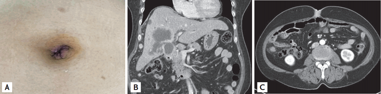
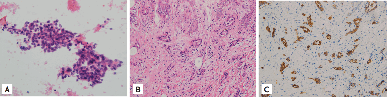
A 61-year-old woman presented to the hospital with a 2-month history of a tender nodule on the umbilicus and an 1-week history of icteric sclera. Her vital signs were within normal ranges. On physical examination, we noted an 1-cm sized, hard and painful nodule on the umbilicus without swelling (Fig. 1A). The laboratory test showed the following findings: hemoglobin 11.4 g/dL, aspartate aminotransferase 123 U/L, alanine transaminase 74 U/L, total bilirubin 12.98 mg/dL, C-reactive protein 2.29 mg/dL, and alkaline phosphatase 477 IU/L. Cancer antigen 19-9 level was 3,880 U/mL. Abdominopelvic computed tomography showed a 7.4 ├Ś 5.9 ├Ś 5.4 cm sized intrahepatic mass arising from the fundus of the gallbladder, multiple enlarged abdominal lymph nodes, enhancing seeding nodules in the peritoneum and focal umbilical thickening with enhancement (Fig. 1B and 1C). Ultrasound-guided needle aspiration cytologic examination of the intrahepatic mass confirmed the diagnosis of adenocarcinoma of the gallbladder (Fig. 2A). The result of skin biopsy of the umbilicus was compatible with metastatic adenocarcinoma (Fig. 2B and 2C). Her malignancy was judged to be inoperable due to the extent of the disease. A biliary stent was inserted to relieve jaundice. Palliative chemotherapy (cisplatin + gemcitabine) every 3 weeks was started.
Sister Mary Joseph nodule, an umbilical metastatic nodule, is reportedly present in 1% to 3% of all intra-abdominal and/or pelvic malignancies. The most common primary source is gastric carcinoma in men and ovarian carcinoma in women. Although Sister Mary Joseph nodule is unusual in a cancerous condition, this case suggests that Sister Mary Joseph nodule can be a crucial initial manifestation of widespread gallbladder cancer, and therefore, careful physical examination is necessary.
 PDF Links
 PubReader
 ePub Link
 Full text via DOI
 Download Citation
 Print
Print


