


To the Editor,
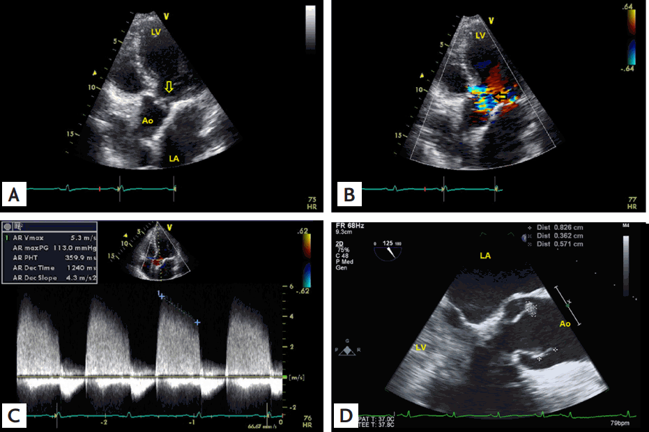
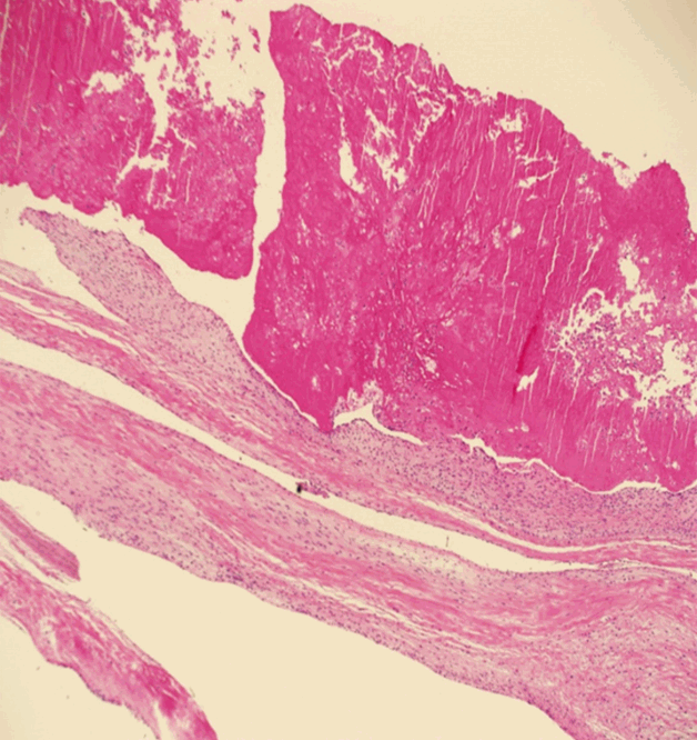
A 53-year-old man was admitted because of fever and right upper quadrant pain that had persisted for 5 days. He had no previously known disease. His initial temperature was 38.3┬░C, and a grade III diastolic murmur was auscultated at the right upper sternal border. The initial laboratory findings were suggestive of acute promyelocytic leukemia and a disseminated intravascular coagulation (DIC) state was suspected. The levels of the inflammatory markers such as the high-sensitivity C-reactive protein (9.6 mg/dL) and procalcitonin (0.774 ng/mL) were slightly elevated. Contrast-enhanced abdominal computed tomography revealed multiple embolic infarctions in the spleen and kidney. Transthoracic echocardiography revealed an echogenic mass attached to the aortic valve (AV) (Fig. 1A), and Doppler studies revealed moderate to severe aortic regurgitation (AR) (Fig. 1B and 1C). Further evaluation with transesophageal echocardiography revealed low echogenic masses attached to the tip of the right coronary cusp and noncoronary cusp (Fig. 1D), resulting in moderate to severe AR. Based on the aforementioned findings, he was highly suspected as having infective endocarditis involving the AV. Because he was in a neutropenic state and had fever, we started intravenous antibiotics (vancomycin and gentamicin) immediately after obtaining blood cultures. Despite using broad-spectrum antibiotics, follow-up echocardiography performed after 1 week revealed no shrinkage of the vegetations and persistent moderate to severe AR. Moreover, recurrent fever was noted. Therefore, the possibility of local uncontrolled infection could not be ruled out. Although blood sample cultures yielded negative results, the patient was scheduled to undergo AV replacement and removal of the infection focus, given that the possibility of culture-negative infective endocarditis by fastidious organisms still existed in this clinical setting (febrile immune compromised patient who is scheduled to receive high dose chemotherapy). Moreover, the patient had developed thromboembolic events. The operative findings were consistent with the preoperative findings, and his postoperative course was uneventful. However, the final pathological examination showed that the vegetation adhered to the valve endothelium was mainly composed of fibrin and had few inflammatory cells (Fig. 2). The most important pathologic finding was a lack of inflammatory response. Therefore, he was diagnosed with nonbacterial thrombotic endocarditis (NBTE), and chemotherapy (arsenic trioxide combined with all-transretinoic acid) was started. He continued to do well clinically without fever or dyspnea.
Clinical diagnosis is quite tricky when vegetations are combined with fever in an autoimmune or myeloproliferative disease because of the vegetation sites being identical to those in infective endocarditis [1]. Furthermore, fever is a common presentation of diseases that are of either metabolic or infective etiologies [1]. Additionally, infective endocarditis by blood culture-negative fastidious organisms should also be considered [2,3]. However, as in the present case, valve replacement could be considered in patients with combined valve dysfunction, and histological evaluation of the resected valve tissue may facilitate the diagnosis [2].
In conclusion, despite the low incidence of NBTE, when cultures yield negative results and highly thrombogenic patients are not responsive to empirical antibiotic treatment, the possibility of NBTE should be considered.
 PDF Links
 PubReader
 ePub Link
 Full text via DOI
 Download Citation
 Print
Print


