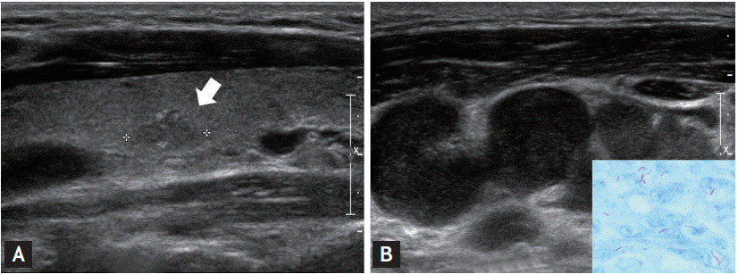
A 31-year-old man was admitted to the outpatient department with an enlarged neck mass that had been growing gradually over the previous month. His medical history was unremarkable, but his mother received anti-tuberculosis medication due to cervical tuberculous lymphadenopathy 10 years ago. Ultrasonography revealed an irregular hypoechoic nodule with calcifications, measuring 1 cm in diameter in the thyroid and multiple enlarged lymph nodes along the jugular chain of the neck. A malignant thyroid nodule with either Kikuchi disease, tuberculosis or metastasis from other lymph nodes was suspected (Fig. 1).
Fine needle aspiration cytology was performed. The thyroid nodule was suspicious for papillary thyroid carcinoma (PTC). Needle biopsy of lymph node revealed chronic granulomatous inflammation consistent with tuberculosis. Upon Ziehl Neelsen carbo- fuchsin staining, a few acid-fast bacilli were identified. Nested polymerase chain reaction (PCR) amplifications were performed using Mycobacterium tuberculosis IS6110 primers (MTBPCR kit, Biosewoom, Seoul, Korea). PCR detected Mycobacterium tuberculosis (Fig. 1B, inset). Chest X-rays were unremarkable. Tuberculosis culture was not performed. An anti-tuberculosis HERZ (isoniazid [INH], rifampin [RFP], ethambutol [EMB], and pyrazinamide [PZA]) regimen was started and continued for 6 months.
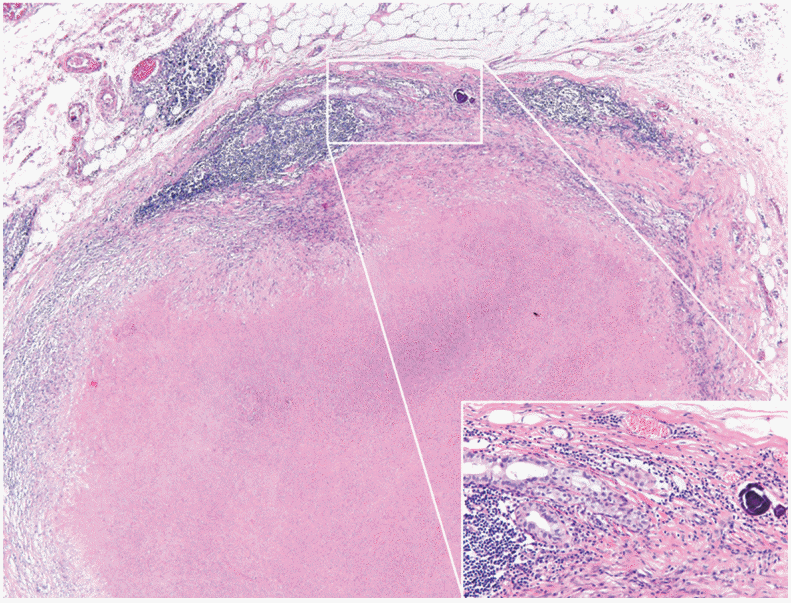
Computerized tomography was performed after starting medication, and it revealed few lymph nodes that remained swollen. Subsequently, a right hemithyroidectomy with neck dissection was performed and was confirmed as PTC. Intriguingly, metastatic PTC was associated with chronic granulomatous inflammation in a single lymph node. Upon whole view examination of the lymph node, lymph node structures were seen at the periphery of the metastatic PTC (Fig. 2). At 2-month postoperative follow-up visit, no complications were observed, and the patient remained well.
Generally, cervical lymphadenopathy originates from metastasis or infection, and metastasis and infection have been identified as main causative factors for lymphadenopathy. The simultaneous occurrence of tuberculosis and metastatic carcinoma is unusual without pulmonary or thyroid tuberculosis. Synchronous dual occurrence of tuberculosis and metastatic carcinoma is often confusing for clinicians, because its presentation is rare. When lymph nodes are continuously enlarged in spite of receiving anti-tuberculosis therapy, the possibility of coexistent metastasis or other malignancies shall not be overlooked.


 PDF Links
 PubReader
 ePub Link
 Full text via DOI
 Download Citation
 Print
Print


