


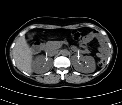
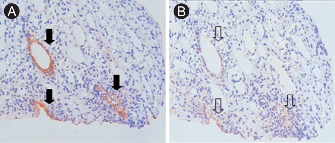
A 44-year-old female was referred to our outpatient clinic with metabolic acidosis and vomiting. She had a history of toxic hepatitis, but no medications. On admission, her blood pressure was 120/70 mmHg with a pulse of 68 beats/min. The laboratory findings showed normal anion gap metabolic acidosis: Na 140 mmol/L, K 1.4 mmol/L, Cl 113 mmol/L, pH 7.192, and bicarbonate 12 mmol/L. The urine chemistry was pH 7.0, Na 55 mmol/L, K 14 mmol/L, and Cl 63 mmol/L (hyperchloremic metabolic acidosis of renal origin). Abdominal computed tomography revealed medullary nephrocalcinosis (Fig. 1). The renal pathology was non-specific. There was decreased net H+ secretion in the collecting tubules, resulting in a high urine pH. Acidification in the collecting tubules is primarily achieved via H secretion by luminal H+-ATPase. However, immunostaining for H+-ATPase was negative (Fig. 2). After alkali replacement with sodium bicarbonate, the serum bicarbonate recovered to above the lower normal limit (26.6 mmol/L). She was discharged in an improved condition.
 PDF Links
 PubReader
 ePub Link
 Full text via DOI
 Download Citation
 Print
Print


 |
 |